One of the things that pushed me toward neuroscience was the desire to understand memory and how we learn things. I know many others who feel the same way, but I doubt any of us really appreciated how difficult a task this would be. A complete picture of how memory works is still out of our reach, but scientists from the University of Leicester and the University of California, Los Angeles recently pushed our understanding a bit further.
The group, led by Drs. Matias Ison, Rodrigo Quian Quiroga, and Itzhak Fried, discovered that individual neurons have the ability to rapidly alter their activity as we learn to associate things [1] . This research provides a framework for understanding what neurons might do as we begin to form a memory, especially of one-time events that occur throughout the day.
To really grasp this experiment’s findings, it helps to understand what the researchers actually did. Epileptic patients undergoing neurosurgery volunteered to participate in the experiment. To find out where their seizures were, researchers implanted single or multi-unit electrodes in patients’ brains. These electrodes also allowed them to record neural activity while showing them different pictures. Recordings were done in the medial temporal lobe, which contains regions like the hippocampus, entorhinal cortex, and amygdala – all locations associated with memory formation and/or processing.
After implantation of the electrode, but before the actual experiment, patients were shown two classes of images: one of people or animals (mainly people, as they caused more neural activity than animals), and the other of famous places like the Eiffel Tower or the White House. Researchers identified which pictures evoked the most activity, calling this the “preferred stimulus”. They then combined the preferred stimulus with a non-preferred stimulus – a picture of the other class that did not cause a change in baseline activity during these pre-experiment trials. In other words, if a neuron was particularly active when shown an image of a cat, but not the Eiffel Tower, the cat might be paired with the Eiffel Tower for the next round of testing.
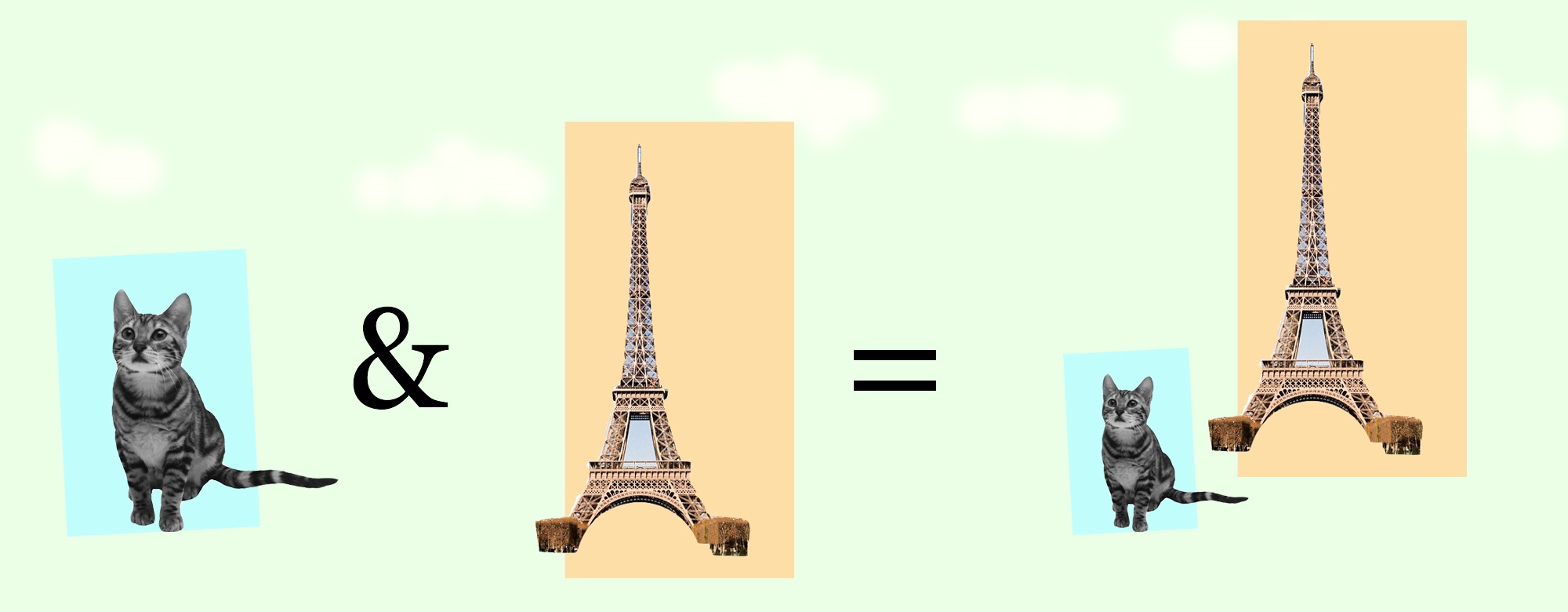
Illustration of the types of images patients might be shown. After assessing neural responses, a preferred image, perhaps a cat, would be paired with a non-preferred image, like the Eiffel Tower.
Throughout the course of their experiments, however, the researchers found that after showing the images containing both the preferred and non-preferred stimuli patients learned to associate the two very quickly in a behavioral task. Moreover, this behavior was accompanied by the neuron (or group of neurons) firing at rates much closer to that of the preferred stimulus when shown either the preferred, composite, or non-preferred image, but not others.
Below are data showing the neural activity and behavioral activity. The left image shows the activity on a trial-by-trial basis, and the right image shows the same activity rearranged for each individual based on the time of learning. In the right graph, point zero is the trial where learning occurred, regardless of when that trial actually was for that patient. So, if neurons fired at similar, elevated rates after one trial for Patient A, this would be point 0 for Patient A. But if it took three trials for Patient B, their data would be adjusted so point zero began at their third trial.
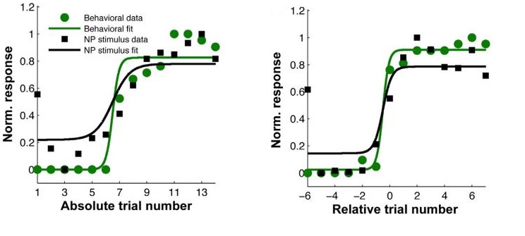
Graphs from Ison, Quiroga, and Fried. Both compare behavioral (green) and neural (black) responses. The graphs show the same data, but the figure on the left has re-aligned the data so that the learning trial is “Relative trial number” 0 for all patients. This accounts for the variability in learning time for different individuals.
This re-arrangement was done to illustrate that when accounting for variability in the time it takes different individuals to learn the associations, there is a strong similarity between a correct behavioral outcome – that is, making the proper association – and increases in neural activity rates. It would be an interesting addition, however, to show individual data for patients learning at different rates to see if they still had similar neural activity learning curves. Such graphs would give a better idea of whether or not learning was always accompanied by a sharp increase in firing rate, or if it were a more gradual process, requiring multiple trials in some individuals. These features are lost in the population data presented here.
The general idea behind this research, that specific neurons are involved with the perception of specific things, is not new. In his 1949 work T he Organization of Behavior , famous neuroscientist Donald Hebb postulated that “a particular perception depends on the excitation of particular cells at some point in the central nervous system” [2] . The idea has also been experimentally confirmed many times with the observation that certain neurons in distinct regions of the brain can be “tuned” to a particular sensory stimulus, like a neuron in the visual cortex that is only active when something of a specific orientation enters the visual field.
It has also been shown before that single neurons can alter their activity as they learn to make associations. When monkeys are shown a complex visual stimulus and rewarded for looking in a certain direction, some neurons in the hippocampus increase their firing rate as the association between the correct direction and reward is learned [3] .
What the recent study offers is the finding that neurons tuned to a particular preferred stimulus can widen their tuning to include other stimuli with which an association is learned. Though this is similar to Wirth et. al.’s finding in monkeys, the authors of this study claim that the increase in activity is the result of changes in a single neuron. Wirth et. al. suggest that other neurons are likely recruited to increase activity as the association forms.
https://greymattersjournal.org/content/images/2015/07/mmc2.mp4
Most strikingly, there is some evidence that the changes in firing pattern, which the authors equate with learning, can happen very quickly. In the learning task – where patients were shown composite images of preferred and non-preferred stimuli – it only took, on average, 3 trials for individuals to meet the criteria for learning. Some, the authors claim, made the association after one trial. As seen above, this was accompanied by changes in neural activity which, supposedly, occurred as the association was encoded.
This research provides an idea of what’s going on at the neuronal level as we learn associations. It also suggests that these changes might be occurring to enable us to remember things that only happen once. So, although this research alone does not show that associations are made because neurons alter their firing patterns, it does provide empirical evidence that can be used to further our theories for how memories, particularly associations, are created, and how neurons behave while forming them.
- M. J. Ison, R. Quian Quiroga, I. Fried. Rapid encoding of new memories by individual neurons in the human brain. Neuron. Vol. 87, Issue 1. 1 July, 2015. Pages 220 – 230.
- D. O. Hebb, The Organization of Behavior. Wiley, New York, 1949.
- S. Wirth, M. Yanike, L.M. Frank, A.C. Smith, E.N. Brown, W.A. Suzuki. Single neurons in the monkey hippocampus and learning of new associations. Science 6 June 2003 : 300 ( 5625 ), 1578 – 1581 .