
Anyone who has ever flown across the world is aware of the exhausting journey that a 12-hour flight can hold, from headaches due to the engine’s grueling background noise to ears popping from pressure differentials. Now, imagine that journey on a larger scale. Instead of simply being 30,000 feet above the ground, envision being about 240 miles above the Earth’s surface in the International Space Station [1]. The “flight” duration is no longer 12 hours, but a whopping 288 consecutive days [2].
This was the plight of Peggy Whitson, who became the U.S. astronaut with the most accumulated days (665) ever spent in orbit after her 288-day flight ended in 2017 [2]. Studies show that spending time in the microgravity environment of low earth orbit comes with debilitating effects. These effects range from muscle atrophy and blurred vision to a recently discovered phenomenon known as the “floating brain.”
The Neurolab Mission & Previous Research Conducted
Although the “floating brain” finding is a more recent discovery, it is only one of many studies that have been conducted regarding microgravity and the brain. In 1998, a 16-day space mission called Neurolab (STS-90) was launched to study the impact of microgravity on the nervous system [3]. During this mission, the crew members conducted a total of 26 experiments on mice, rats, snails, crickets, two types of fish, and themselves. Tests occurred before, during, and after spaceflight to acutely determine how microgravity affects neural functions. Specifically, Neurolab investigated sensory integration and navigation, blood pressure control, the balance system, circadian rhythms and sleep, and nervous system development in weightlessness [3].
Among many complex, detailed findings, Neurolab revealed that astronauts’ responses to tilting and rolling motions are impacted by microgravity, which has implications for their motor and sensory skills [3]. Another experiment using a virtual environment display system showed that astronauts rely more on visual cues in microgravity conditions than in normal gravity conditions [3]. The experiments clearly indicated that astronauts return to Earth with a nervous system significantly different than the one they left with.
Two other studies demonstrated microgravity’s ability to change rats’ brains. One showed that short- and long-term changes in neural structure are similar to neuronal alterations in conditions such as stroke [3]. A second experiment compared the brains of rats in space with a control group raised on Earth and indicated that rats’ brains developed less in space and younger rats were more likely to die [3].
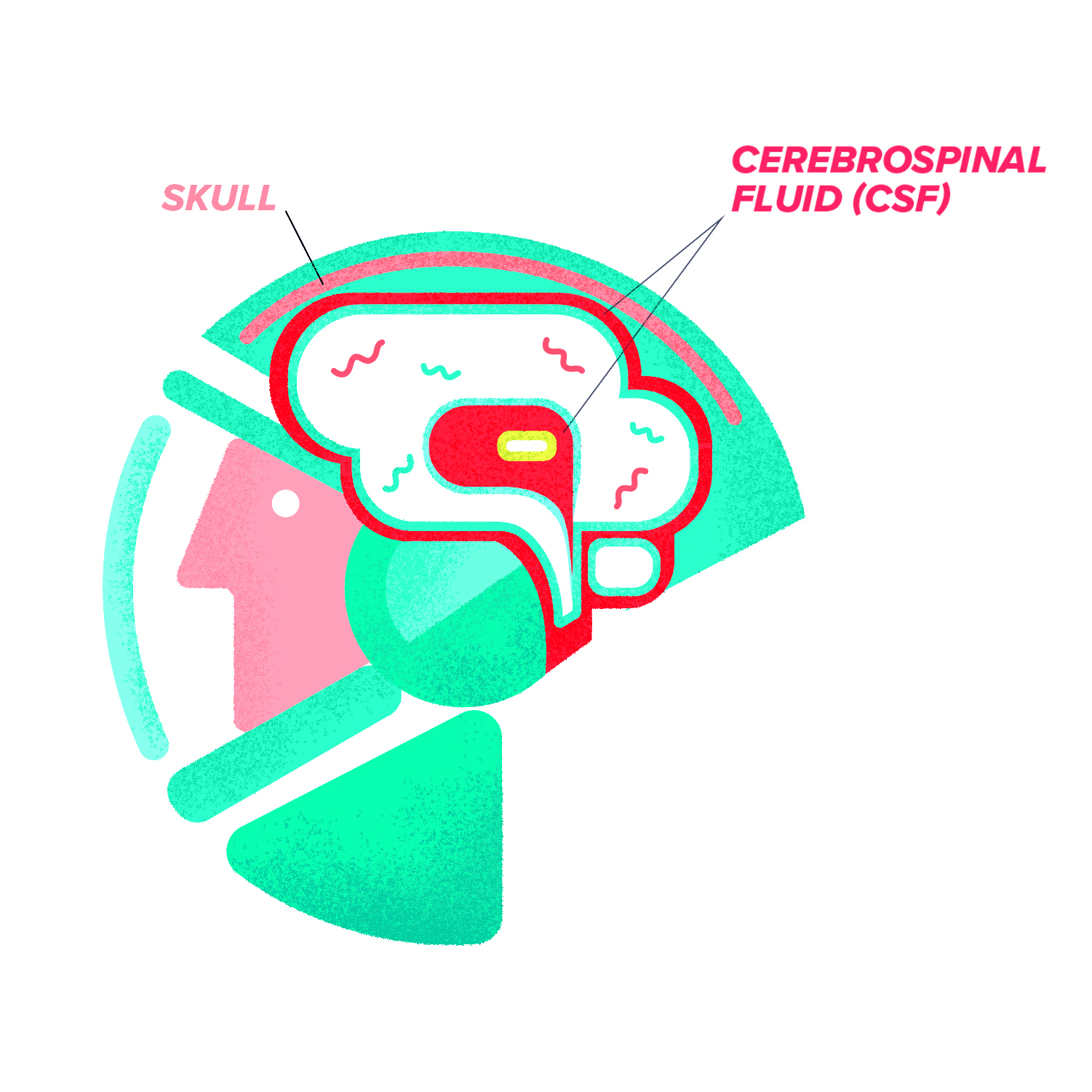
Furthermore, many studies have also investigated the role of microgravity on cerebrospinal fluid (CSF), a colorless, protective liquid that surrounds the brain. CSF circulates nutrients and functions as a shock absorber. A study by Dr. Larry Kramer’s team at the University of Texas Health Science Center investigated the relationship between CSF production and globe flattening, where the eyeball is compressed from its original shape [4]. Dr. Kramer’s team found that astronauts with notable posterior globe flattening had a 70% increase in CSF production rate post-flight. This finding is consistent with the hypothesis that CSF production decreases in the microgravity environment of space but increases when astronauts return to normal gravitational conditions [4]. Likewise, a study conducted by Dr. Yasuaki Kawai’s team of Tottori University found that microgravity causes a fluid shift from the lower body to the upper body [5]. As such, both of these studies support the idea of microgravity affecting CSF distribution.
The “Floating Brain”
The “floating brain” phenomenon is a recent addition to the research exploring the effects of microgravity on the human body [6]. Neuroradiologist Dr. Donna Roberts of the Medical University of South Carolina and other researchers conducted a study comparing magnetic resonance imaging (MRI) scans of astronauts’ brains before and after missions, including 18 astronauts after long missions (about 165 days) and 16 astronauts after short missions (about 14 days). The scientists focused on changes in volume of CSF spaces at the top surface of the brain, vertical displacement of the brain, and volume of the central sulcus [6].
These aspects were important to measure due to the vital roles of CSF and the central sulcus in the brain. Scientists’ currently believe that CSF imbalances can affect the cognitive function of the brain [7]. The central sulcus is an area of the brain that divides the frontal lobe, important for reasoning and speaking, from the parietal lobe, which is important for memory and hearing. Specifically, the central sulcus is the absence of tissue, separating the motor cortex in the frontal lobe from the somatosensory cortex in the parietal lobe. Due to its position as a divider, changes in the central sulcus reflects changes in its surrounding tissues. The surrounding tissues in this case are implicated in motor function, which is especially important for astronauts since they are constantly adapting to new environments to execute their tasks [8].
Roberts and her research team took MRIs of the astronauts’ brains about a week after spaceflight. They found that the central sulcus narrowed significantly for 17 out of 18 astronauts who went on long duration flights, but for astronauts on short duration flights, only three out of 16 had narrowing of the central sulcus [6]. The researchers hypothesized that the central sulcus narrowed due to the increase in volume of the frontal and parietal lobes. Scientists are still exploring how this phenomenon influences motor function. Roberts’ team also analyzed a subgroup of the astronauts and discovered that the brain had shifted closer to the top of the skull for all of the astronauts in the long duration study group, but only half of the astronauts in the short duration study group. In addition, near the upper surface of the brain, CSF spaces decreased for all astronauts on long duration flights. The same effect was observed for only one out of six astronauts on short duration flights [6].
Consequently, the scientists concluded that staying in space for long periods of time causes the brain to shift or “float” upward [6]. While researchers have yet to determine if this effect reverses itself over time, the “floating brain” phenomenon has potentially profound implications. The upward shift of the brain can compress veins or venules, obstructing CSF and venous outflow. As a result, intracranial pressure can increase, resulting in swelling of the optic nerve [6].
Additional Recent Findings
While the “floating brain” study highlights important effects of microgravity on the position of the brain and CSF spaces in the skull, other studies have investigated the impact of space travel on ocular health as well as grey matter and white matter. For instance, spaceflight can have a range of debilitating effects on white matter [9]. White matter is primarily made of neurons’ axons, which connect to other neurons’ dendrites and allow for communication between cells. A study led by Dr. Noam Alperin, professor of biomedical engineering and radiology at Miami University Miller School of Medicine, investigated a correlation between CSF and white matter hyperintensity in astronauts [9]. White matter hyperintensity refers to white matter areas that appear lighter in color on MRIs than surrounding tissues due to axonal degeneration within neurons [10]. In general, increased white matter hyperintensity is linked to cognitive impairment [10]. The scientists found that only astronauts on long-duration flights had an increase in white matter hyperintensity around ventricles of the brain, a connected system of cavities filled with CSF [11]. This increase in white matter hyperintensity was also linked to an increase in ventricular CSF volume, which has been associated with old age, Alzheimer’s disease, and a higher risk of stroke [10][11][12].
Spaceflight can also affect grey matter. Grey matter contains neuronal cell bodies and dendrites, which process signals from other cells before transmitting the signaled response. Dr. Vincent Koppelmans of the University of Utah and colleagues found huge volumetric decreases of grey matter in the temporal lobe, which is vital for auditory processing, as well as in the frontal lobe, which controls executive function among many other complex physiological processes [13]. Koppelmans hypothesized that grey matter was redistributed in response to microgravity and other stressors during an astronaut’s mission. However, Koppelmans and his team also noticed grey matter increases in the motor cortex. The researchers hypothesized that the stressors of space travel, like sleep loss, could result in brain atrophy [13].
Recent studies have also indicated that spaceflight can impact ocular health, which could possibly be a result of changing the space that CSF occupies, affecting the optic nerve. 29% of astronauts on short duration flights and 60% of astronauts on long duration flights developed visual deficiencies that did not heal for years after spaceflight [14].
Specifically, Dr. Noam Alperin and his team observed globe flattening in the eyes of astronauts, which could lead to farsightedness, a phenomenon that astronauts on the International Space Station often experience [15]. Alperin’s team also observed a correlation between increased orbital CSF volume and ocular changes. Normally when people stand up, CSF gets pulled from the brain into the spinal cord by gravity. In space, the lack of gravity enables the movement of CSF into the eye socket [15].
Additionally, a recent study conducted on twin astronauts aims to further outline the impact of microgravity on the human body. Astronaut Scott Kelly went into space for a year, while his identical twin brother Mark Kelly remained on Earth [18]. Preliminary findings have revealed some physiological and cognitive changes. For instance, Scott Kelly showed a decrease in the speed and accuracy of his cognitive performance post-flight compared to Mark Kelly [18].

What Does This Mean for NASA?
When astronauts are chosen for missions, they undergo extensive health screenings and training to assess how they will endure the stressors that come with the microgravity environment of space. NASA also ensures that astronauts perform health screenings during spaceflight. In fact, during Peggy Whitson’s 288-day journey, she used a device called a fundoscope that monitored her ocular health by examining interior eye structures, such as the retina [2][19].
In the future, NASA is considering sending astronauts to Mars, a flight that could take more than 300 days, making it considerably longer than most current missions [2]. The findings from the Neurolab mission indicated that NASA should consider the relationship between gravity and human development, as there are periods of time when gravity is critical to development [13]. Moreover, Dr. Donna Roberts and her team from the “floating brain” study believe that NASA’s imaging protocols should include advanced MRI techniques and repeated imaging of the brain over a longer period of time after spaceflight [6].
Consequently, NASA should consider making more changes to future space shuttles to counteract microgravity, preventing potential long-term effects on the brain. Each new research study is a small “step” toward a better understanding of the brain in space. However, each “leap” forward for mankind by astronauts’ research during space missions should not come at a cost to the brain and nervous system.
References
- Thirsk, R., Kuipers, A., Mukai, C., & Williams, D. (2009). The space-flight environment: the International Space Station and beyond. Canadian Medical Association Journal, 180 (12), 1216-1220. doi:10.1503/cmaj.081125
- Astronaut Biography: Peggy A. Whitson. (2017, September). NASA. Retrieved February 12, 2018.
- Buckey, J. C., & Homick, J. L. (Eds.). (2003). The Neurolab Spacelab mission: neuroscience research in space: results from the STS-90, Neurolab Spacelab mission . Houston, TX: National Aeronautics and Space Administration, Lyndon B. Johnson Space Center.
- Kramer, L. A., Hasan, K. M., Sargsyan, A. E., Wolinsky, J. S., Hamilton, D. R., Riascos, R. F., . . . Otto, C. (2015). MR-Derived Cerebral Spinal Fluid Hydrodynamics as a Marker and a Risk Factor for Intracranial Hypertension in Astronauts Exposed to Microgravity. Journal of Magnetic Resonance Imaging,42 (6), 1560-1571. doi:10.1002/jmri.24923
- Kawai, Y., Doi, M., Akira, S., Reiko, H., Keigo, U., Yasumasa, A., & Kyoko, T. (2003). Effects of Microgravity on Cerebral Hemodynamics. Yonago Acta Medica,46 (1), 1-8. Retrieved March 5, 2018.
- Roberts, D. R., Albrecht, M. H., Collins, H. R., Asemani, D., Chatterjee, A. R., Spampinato, M. V., . . . Antonucci, M. U. (2017). Effects of Spaceflight on Astronaut Brain Structure as Indicated on MRI. New England Journal of Medicine,377 (18), 1746-1753. doi:10.1056/nejmoa1705129
- Sakka, L., Coll, G., & Chazal, J. (2011). Anatomy and physiology of cerebrospinal fluid. European Annals of Otorhinolaryngology, Head and Neck Disease,128 (6), 309-316. doi:10.1016/j.anorl.2011.03.002
- Petrides, M. (2014). Neuroanatomy of Language Regions of the Human Brain. doi:10.1016/B978-0-12-405514-8.50006-2
- Alperin, N., Bagci, A. M., & Lee, S. H. (2017). Spaceflight-induced changes in white matter hyperintensity burden in astronauts. Neurology,89 (21), 2187-2191. doi:10.1212/wnl.0000000000004475
- Wardlaw, J. M., Valdés Hernández, M. C., & Muñoz-Maniega, S. (2016). What are White Matter Hyperintensities Made of?: Relevance to Vascular Cognitive Impairment. Journal of the American Heart Association,5 (1). doi:10.1161/jaha.115.002006
- Crisan, E., MD, Jasvinder, C., & Salim, B. R. (2016). Ventricles of the Brain. Medscape. Retrieved February 12, 2018.
- Ott, B. R., Cohen, R. A., Gongvatana, A., Okonkwo, O. C., Johanson, C. E., Stopa, E. G., . . . Silverberg, G. D. (2010). Brain Ventricular Volume and Cerebrospinal Fluid Biomarkers of Alzheimers Disease. Journal of Alzheimers Disease , 20(2), 647-657. doi:10.3233/jad-2010-1406
- Koppelmans, V., Bloomberg, J. J., Mulavara, A. P., & Seidler, R. D. (2016). Brain structural plasticity with spaceflight. Npj Microgravity,3 (1). doi:10.1038/s41526-016-0001-9
- Lee, A. G., Tarver, W. J., Mader, T. H., Gibson, C. R., Hart, S. F., & Otto, C. A. (2016). Neuro-Ophthalmology of Space Flight. Journal of Neuro-Ophthalmology,36 (1), 85-91. doi:10.1097/wno.0000000000000334
- Alperin, N., Bagci, A. M., Oliu, C. J., Lee, S. H., & Lam, B. L. (2017). Role of Cerebrospinal Fluid in Spaceflight-induced Ocular Changes and Visual Impairment in Astronauts. Radiology . doi:10.1148/radiol.2017161981
- Benvenuti, S. M., Bianchin, M., & Angrilli, A. (2011). Effects of Simulated Microgravity on Brain Plasticity: A Startle Reflex Habituation Study. Physiology & Behavior,104 (3), 503-506. doi:10.1016/j.physbeh.2011.05.019
- Kramer, L. A., Sargsyan, A. E., Hasan, K. M., Polk, J. D., & Hamilton, D. R. (2012). Orbital and Intracranial Effects of Microgravity: Findings at 3-T MR Imaging. Radiology,263 (3), 819-827. doi:10.1148/radiol.12111986
- Edwards, M., & Abadie, L. (2018, January 31). NASA Twins Study Confirms Preliminary Findings. NASA . Retrieved March 5, 2018.
- Norsk, P., Otto, C., Barr, Y., Schelhamer, M., & Davis, J. (2015). The Visual Impairment Intracranial Pressure Syndrome in Long Duration NASA Astronauts: An Integrated Approach. NASA. Retrieved February 12, 2018.