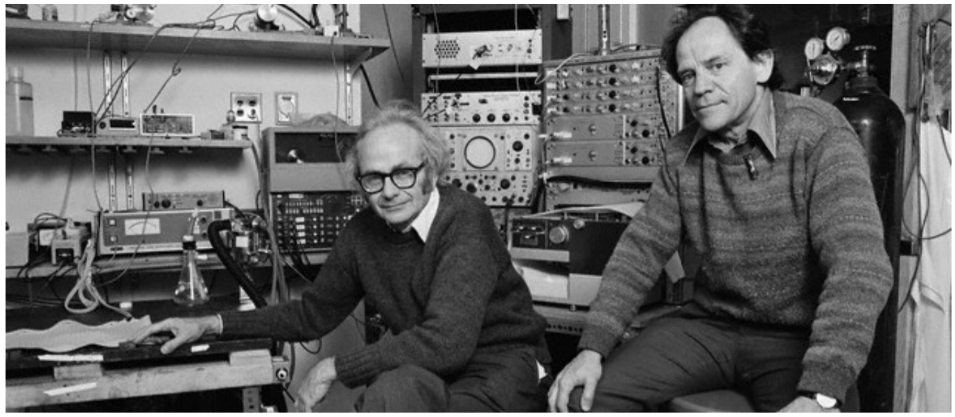
The first step in any research endeavor is to start with the basics. David Hubel and Torsten Wiesel were some of the pioneers of sight research who focused on the monumental task of studying the basic processing and architecture of the visual system.
Humans see the world as a united and complete picture. However, the visual system is actually surprisingly specialized at a cellular level [1]. This was partially revealed by the research of Hubel and Wiesel in a series of experiments and papers produced from the 1950’s to the 1980’s [1].
Their findings were impactful enough that in 1981, they shared the Nobel Prize in Physiology. Today, they are considered pioneers of vision research [2].
Retinal-geniculate-striate System
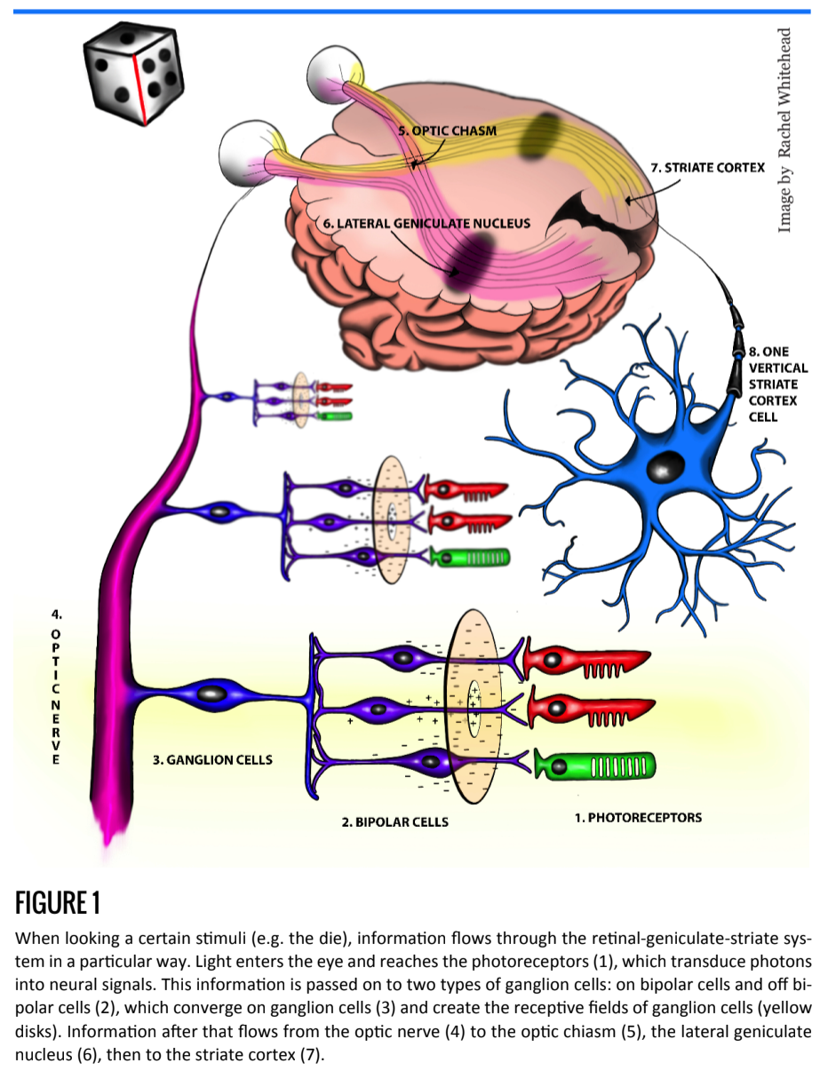
Hubel and Wiesel focused their research on a series of cells known as the retina-geniculate-striate system. The basic flow of information along this pathway, as implied its name, is from the retina to the lateral geniculate nucleus to the striate cortex, also known as the primary visual cortex [3][4] (Figure 1). Within the retina, photoreceptors transduce light energy into neural signals, which are passed first to bipolar cells, then to ganglion cells. These ganglion cells, then code the intensity and duration of the stimulus and project their axons into the optic nerve [4].
The retinal bipolar cells that synapse on the photoreceptors respond to glutamate, and therefore light, in different ways 6. The two types of retinal bipolar cells are called on bipolar cells, which are excited by light, and off bipolar cells, which are inhibited by light [6]. These cells synapse on ganglion cells, where the inhibition and excitation signals compete to cause selective firing of ganglion cells [6]. As previously mentioned, this information follows a specific route to the striate cortex, where processing begins.
Receptive Fields
Hubel and Wiesel were interested in the receptive fields of neurons in the striate cortex [1]. Receptive fields are areas of the vision that activate or inhibit a single neuron in the visual system [4]. Earlier research by Dr. Stephen Kuffler showed that the receptive fields of ganglion cells have both inhibitory and excitatory regions in concentric patterns [7]. In other words: a receptive field has two circular areas, one excitatory region encircled by a larger inhibitory region.
If light shines on the excitatory region, ganglion cells depolarize and fire action potentials. If, however, light stimulates the inhibitory region, they are less likely to do so [7].
The location and pattern of the excitatory region of receptive fields can vary and determines ganglion cell function7. For example, “on center” ganglion cells, shown in Figure 1, best respond to light falling only in the center of the receptive field (the excitatory region). These ganglion cells do not respond well to diffuse light, which causes the excitatory and inhibitory regions of the receptive field to cancel and results in no further signal [7].
Based on these observations, it was inferred that ganglion cells provide information regarding contrast [7].
Hubel and Wiesel expanded Kuffler’s research by focusing on cells within the striate cortex with similar specificity [1].
Research and Discoveries
Although Hubel and Wiesel’s discoveries were monumental, they had a relatively simple experimental design. Using an anesthetized cat with paralyzed eye muscles, the researchers would place microelectrodes near individual visual cortex neurons. With such a set up, they could apply a stimulus (light) to different areas of the visual field until the corresponding cortical neurons fired. In this way, they could locate the receptive field of a visual neuron in the striate cortex.
Having found a specific receptive field, they experimented with placement and shape of light in that receptive field to determine the areas of excitation and inhibition [8]. They found, for example, that some neurons responded optimally to bars of light in a specific orientation but not another [8] (e.g. vertical light vs. horizontal light). These cells became known as simple cells [1].
Hubel and Wiesel hypothesized that the receptive fields of simple cells were formed in an additive fashion as ganglion cells and their receptive fields converged in the lateral geniculate nucleus [1] (Figure 1.6). Cells with receptive fields that were not easily explained via the addition of retinal ganglion receptive fields became known as complex cells [1].
Hubel and Wiesel further discovered that cells with similar orientation preferences existed in organized columns, called orientation columns, within the striate cortex [10]. Likewise, they discovered ocular dominance columns, that preferentially respond to input from one eye, and are responsible for determining depth perception [1][10].
Further Research and Impact
In follow up work, some researchers have argued in favor of using natural lighting over artificial bars of light to study vision because the nervous system operates in scenes, not in specific pieces [12]. New studies are showing that, in some cases, neurons are inhibited in response to natural stimuli, but excited when exposed to artificial stimuli [12]. Therefore, natural stimuli may better reveal the correct functional pathways.
Additionally, some researchers are exploring how context affects the firing pattern of visual neurons [13]. It has been shown that stimuli placed outside of the observed receptive field in the preferred orientation of that receptive field can actually inhibit the neuron [13]. This may indicate that receptive fields in the visual system are larger than originally thought [13].
Continuing research on simple and complex cells, ocular dominance columns, and orientation columns shows that Hubel and Wiesel’s influence has not faded over time. Their discoveries were monumental in describing the processing of the visual system, helping to clarify its structure, and giving future researchers a foundation upon which to build [14]. There is no doubt that in the future scientists will continue to use Hubel and Wiesel’s groundbreaking work to expand upon the visual system and solve its remaining mysteries.