The Concussion that Caused a Commotion: A Look at King Henry II of France
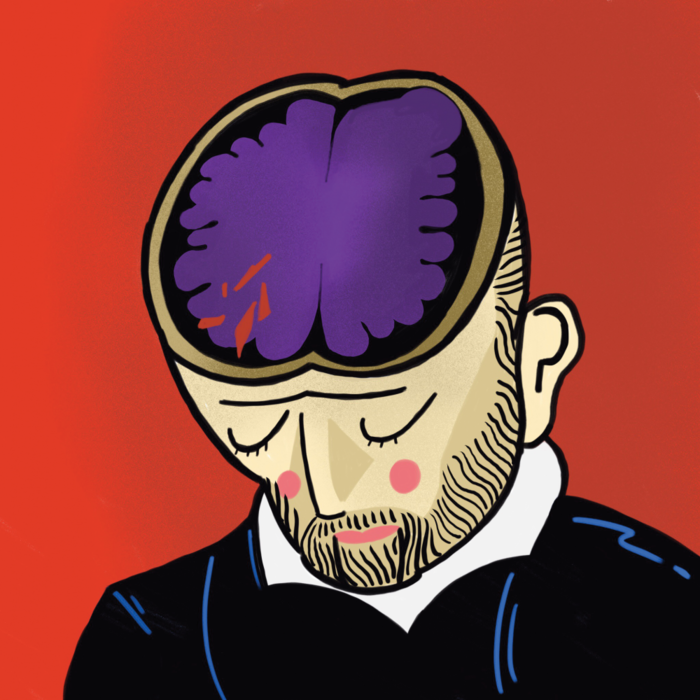
Clad in armor and lances at hand, two jousters mounted their steeds. The competitors aimed their weapons and rushed towards each other on horseback. It was in that moment that one of the jousters was met face on with his opponent’s lance. The wooden lance entered through his faceguard and shattered into fragments, piercing his right eye and penetrating the right orbit and temple of his skull. The most scandalous part of this story is that the jouster who received the blow happened to be the king of France! On June 30, 1559, during the royal wedding of his daughter Elisabeth and King Philip II of Spain, King Henry II of France was struck in the face with a jousting stick by Count Gabriel de Montgomery [1] . It made for quite the shock when the king, still conscious, picked himself up after falling from the strike. He was escorted into the palace immediately for care. What followed during the treatment of King Henry II of France laid the foundation for how we understand concussions and the effects of traumatic brain injuries today.
Observations of the King
King Henry II of France was born in 1519 and reigned from 1547 until 1559, when he succumbed to his injuries eleven days after the jousting accident [3] . Although Henry was left blinded in the right eye, the initial head injury did not immediately affect the king and His Majesty went on fulfilling his kingly duties. He was fully alert throughout his initial treatment [2] . The night of the injury, many of the king’s physicians came to his bedside to extract multiple splinters lodged in his forehead [3] .
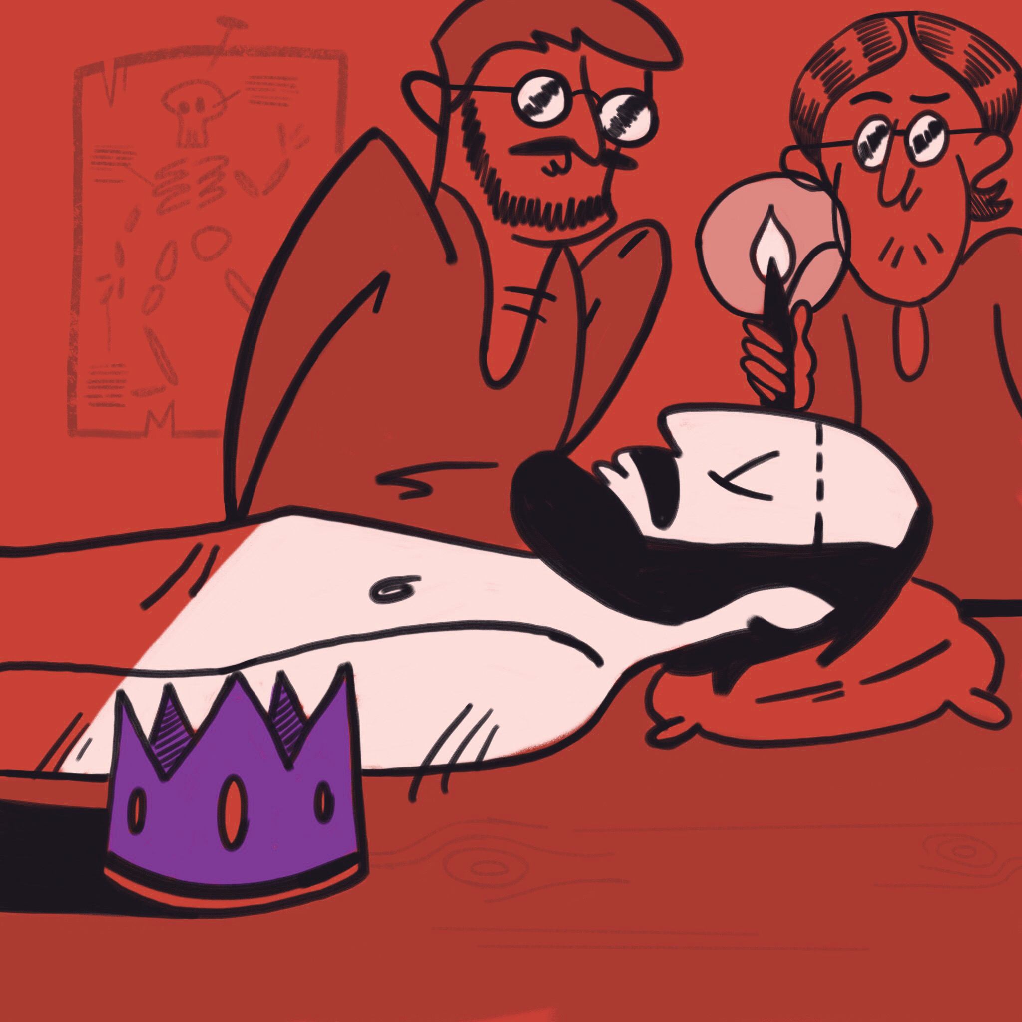
The two royal surgical physicians summoned to the scene to oversee Henry’s care were Ambroise Paré and Andreas Vesalius. Ambroise Paré served as King Henry’s royal physician and was experienced in surgical treatment and battlefield medicine. Vesalius was an anatomist who served King Philip II of Spain and was renowned for his publication of the anatomy book, De Humani Corporis Fabrica . These two surgical physicians were tasked to diagnose and treat the injured king.
In one diagnostic experiment, Vesalius instructed the king to bite on a cloth and then pulled the cloth from the king’s mouth. Henry reacted to the pain and threw his hand to his head [3] . The king’s discomfort was a significant clue that more of the cranial region was affected than just his right orbit, or the eye socket. It is implied from accounts and historical reviews, though vague, that the result of the experiment also led Vesalius to predict that the king’s wounds would continue to deteriorate [2] .
After manually removing many splinters that were stuck in the king’s right eye socket, the physicians left the rest of the smaller splinters alone because the king shouted when they tried touching them. Given the fear of inciting further harm to the orbit by performing surgery on the king, Paré and Vesalius chose instead to observe the wound and the health of the king.
On the fourth day of his treatment, the king’s fever rose [2] . On July 9th, Henry experienced left-sided paralysis of his leg and arm and convulsions on the right side of his body. Vesalius reported that his face swelled, his neck stiffened, and he felt throbbing at the back of his skull [1] . Simul-taneously, pus emanated from his right eye socket [1] . Henry’s vision in his left eye blurred, and his headaches continued to increase.
Jousting Experiment
Midway through the king’s care, Paré, Vesalius, and fellow physicians decided to determine the effects of the injury and simulated the jousting accident by experimenting on the decapitated heads of executed criminals [2] . The incident was replicated with a volunteer jouster charging on horseback and aiming his lance at the right eye of the heads [4] . The experiments tested to see if the lance could penetrate the orbit of the decapitated head with a cross-body blow. The results revealed that none of the skulls of the decapitated heads were fractured. The splinters from the jousting sticks had not pierced through the skulls but instead remained in the orbit, suggesting that the jousting stick did not directly damage the brain [1] . This evidence led to the conclusion that Henry had no skull fracture and many of the splinters remained in his eye socket. The results of the experiment were viewed as a sure sign that because King Henry’s skull wasn’t fractured, his trauma would be survivable. But the conclusions made contradicted Vesalius’ earlier diagnostic findings, which indicated that his survivability was indeed low [2] . This news may have also affected the intensity of the king’s care and minimized the severity of the injury following the experiment.
The king continued to physically deteriorate over the eleven days post-injury and died on July 10, 1559 [2] . The physicians were left wondering about the cause of death, given the results from the tests they had done on the king and the jousting experiment. Despite minimal open wounds and indication of little to no skull fractures, something caused the death of King Henry II of France, and it had to be explored.
A Royal Autopsy
After King Henry’s passing, his wife, Queen Catherine de Medici, made the rare and taboo order for an autopsy to be performed on the late king [3] . During the 16th century especially, autopsies were reserved for criminals [5] . Performing an autopsy on someone of high status – let alone the king of France – was considered undignified and degrading. However, both Vesalius and Paré attended and participated in the king’s autopsy with their own observations.
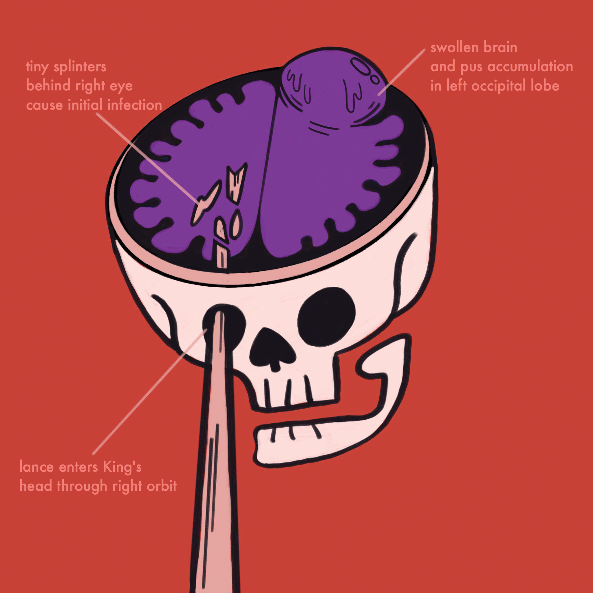
As Vesalius expected during the autopsy, the socket of Henry’s right eye still contained many splinters. While there was no wound exposing the brain, Henry had an infected eye socket from the splinters, which served as an opening for infection to spread through circulation [4] . The left part of Henry’s brain was filled with yellowish fluid that looked affected by putrefaction, or decay of tissue from infection [6] . The infectious lesion was cited to contain pools of blood found at the superficial layer that protects the brain. The damaged regions explain the left-sided paralysis and right-sided convulsions Henry experienced [7] .
Paré added in his own report that most of the pus was at the back of the brain in the left occipital region and far from the wounded right eye. Given current knowledge that the occipital lobe is largely responsible for sight, that damage could also explain the deficit in the vision of the left eye. Paré believed an occipital epidural hematoma was present, a condition that occurs when pools of blood accumulate at the back of the head [3] . As the infection increased over time, at the middle of the occipital bone quantities of blood spread out between the dura and pia layers protecting the brain and formed pus – an accumulation of white blood cells reacting to infection – signifying the onset of putrefaction [3] .
From the observations made by both Vesalius and Paré during the autopsy, the presence of splinters no further than the eye socket matched the results from their earlier simulation. More significantly, the two physicians found a relation between the splinters, the damage to the back of the brain, and the pools of fluids in areas of the brain that was integral to uncovering the effect the jousting accident had on King Henry’s brain. The king’s autopsy also helped normalize autopsies as a standard medical practice for people regardless of social status. The autopsy was essential in revealing what happened to the brain, allowing a better look at the path of the lance’s impact on King Henry’s head and what the impact did to affect the king’s health.
A Controversial Diagnosis
So what exactly killed the king? Was it the infection, or a serious traumatic brain injury? Vesalius and Paré each proposed possible explanations based on their findings from the autopsy.
Due to the pus found in the brain post-mortem, Vesalius concluded that the king had a cerebral abscess, or a collection of pus in the brain. He suggested that the king had developed a severe infection from the tiny splinters in his right eye [1] . As concluded from the jousting experiment, the jousting stick only penetrated the orbit and never directly damaged the brain. However, this still did not account for the damage found at the back of the brain.
Paré entertained the possibility of a concussion that resulted in the king’s death, a violent blow on the brain by the force of the lance [3] . Paré suggested that King Henry’s brain had suffered a contrecoup injury—damage to the brain localized opposite to where the traumatic force originated. In the king’s case, it was a strike at the front which shocked the brain, causing it to hit the back of the skull and damage the occipital lobe. The throbbing in the back of his brain explains Henry’s diminishing vision as well as the putrefying wounds that were present. At times, skull fractures are helpful in relieving a swollen brain by providing additional space [3] . In King Henry’s case, without a fracture to relieve an intracranial injury, the pressure buildup had heightened the damage to the brain.
Modern assessments such as the one made by Zanello suggest that following the initial injury, the king had been afflicted by an initial infection developed at the right orbit because of the wooden splinters, spreading intracranially through facial veins to the site of the hematomas formed at the occipital area [6,8] . The whiplash from the jousting strike had indeed damaged Henry’s brain. His increased headaches suggested that the brain was swelling and the blood vessels inside the skull had probably ruptured, resulting in the infection that caused the convulsions and regression of sight.
Vesalius saw the infections from the splinters as the source of King Henry’s deterioration, whereas Paré considered the sustained injury to the brain from impact to be a critical factor. Though they both held the pieces to uncover what ailed the king after the autopsy, there was no way for them to have used this information to save the king. It proved difficult for the physicians to do anything to help in terms of treatment.
Limited Treatment
Medical science was not prepared to provide a successful treatment to such a complicated and invasive injury at the time [5] . In 16th century Europe, medical practice relied on ideas of the humoral balance, a belief in which the balance of bodily fluids was central to the health and wellness of an individual. The two doctors did not know about neurons or localized pain, and options for treatment were understandably scarce. Making things more difficult for Paré and Vesalius were King Henry’s royal status and the utmost sensitivity that had to be upheld in the king’s care. The two doctors risked charges for malpractice if they were deemed responsible for the king’s death. Although hired under order to provide treatment for the king, the strict pressures contributed to and culminated in serious reservations that affected the king’s prognosis [5] .
Without anesthetics, the splinters were too numerous and small to remove. Exfoliation, a process of washing out foreign bodies via bodily fluids, was believed to be the only way to remove the more unreachable splinters from the king’s wound [4] . Trepanning – when a hole is drilled in the side of the skull – was a common practice to help release fluids, relieve pressure, and ease pain. But this idea was abandoned during the king’s care because of the large quantities of pus already seeping onto the bandages from the eye socket. There was no supposed advantage to trepanning for fluids already seeping out of the body [9] . Regardless, such procedures were often fatal due to risk of neural infection from cutting the dura mater. Before the infection spread to the brain, the two physicians probably did not consider performing an orbital exenteration, a surgical procedure to remove the eye, which later, after further medical advances, was received as an accepted practice in 1583 [9] . Removing the eye may have exposed and elicited more removal of splinters, which could have decreased the spread of infection to the brain and reduced the risk of fatality.
This case brought up questions of how to look at and treat brain injuries. Henry’s case demonstrated how injury to the brain can occur regardless of the presence of a skull fracture. Today, medical imaging has provided a means to make more accurate assessments of the skull and brain following traumatic injury. Modern medicine also provides safer means of extracting foreign objects from sites of injury and administering antibiotics to reduce the severity of infections [1,4] .
Conclusion
King Henry II of France’s condition helps us today in understanding the effects of concussions. The king’s concussion caused quite the commotion not only politically, but also within the scientific community. This story and autopsy in medical history ignited progress in neuroscience. The king’s suffering foreshadowed many great discoveries over the few centuries of neuroscience—it showed how dangerous brain injuries can occur without a fracture and how a concussion does not always lead to immediate unconsciousness [4] . King Henry’s case describes different symptoms not only related to concussions, but also the effects of damage to certain regions of the brain. Modern-day knowledge shows that head trauma can cause blood vessel damage, nerve damage, infection, and seizures, to name a few effects [10] . King Henry’s concussion is echoed today as we continue to learn the long-term effects of concussions on the brain, especially as prevalent as they are in American football and other modern sports. As Vesalius and Paré saw while observing the king of France during his “commotion cérébrale,” there is still much to learn about what can be done to treat trauma and injury to the brain in modern times.
- Eftekhari, Kian. “The last ride of Henry II of France: Orbital injury and a king’s demise.” Comp. Christina Choe, M. Reza Vagefi, and Lauren Eckstein. Survey of Ophthalmology 60.3 (2015): 274-78.
- Norwich I: A consultation between Andreas Vesalius and Ambroise Paré at the deathbed of Henry II. King of France, 15 July 1559 [sic]. South African Med J 80:245–247, 1991
- Zanello, M., Charlier, P., Corns, R. et al. “The death of Henry II, King of France (1519–1559). From myth to medical and historical fact” Acta Neurochirurgica (2015) 157: 145. doi:10.1007/s00701-014-2280-9
- Martin, G. (2001), The death of Henry II of France: A sporting death and post-mortem. ANZ Journal of Surgery, 71: 318–320. doi:10.1046/j.1440-1622.2001.02102.x
- Faria, Miguel A. “The death of Henry II of France.” Journal of Neurosurgery 77.6 (1992)
- C.D. O’Malley. Andreas Vesalius of Brussels 1514–1564. University of California Press, Berkeley and Los Angeles, CA (1964), pp. 283–288 396–8
- Menche N. (ed.) Biologie Anatomie Physiologie. Munich: Urban & Fischer/ Elsevier; 2012.
- Miller CF, Brodkey JS, Colombi BJ (1977) The danger of intracranial wood. Surg Neurol 7(2):95–103
- R.A. Goldberg, J.W. Kim, N. Shorr. Orbital exenteration: results of an individualized approach. Ophthal Plast Reconstr Surg, 19 (2003), pp. 229–236
- Stocchetti N, Zanier ER. Chronic impact of traumatic brain injury on outcome and quality of life: a narrative review. Critical Care. 2016;20:148. doi:10.1186/s13054-016-1318-1.