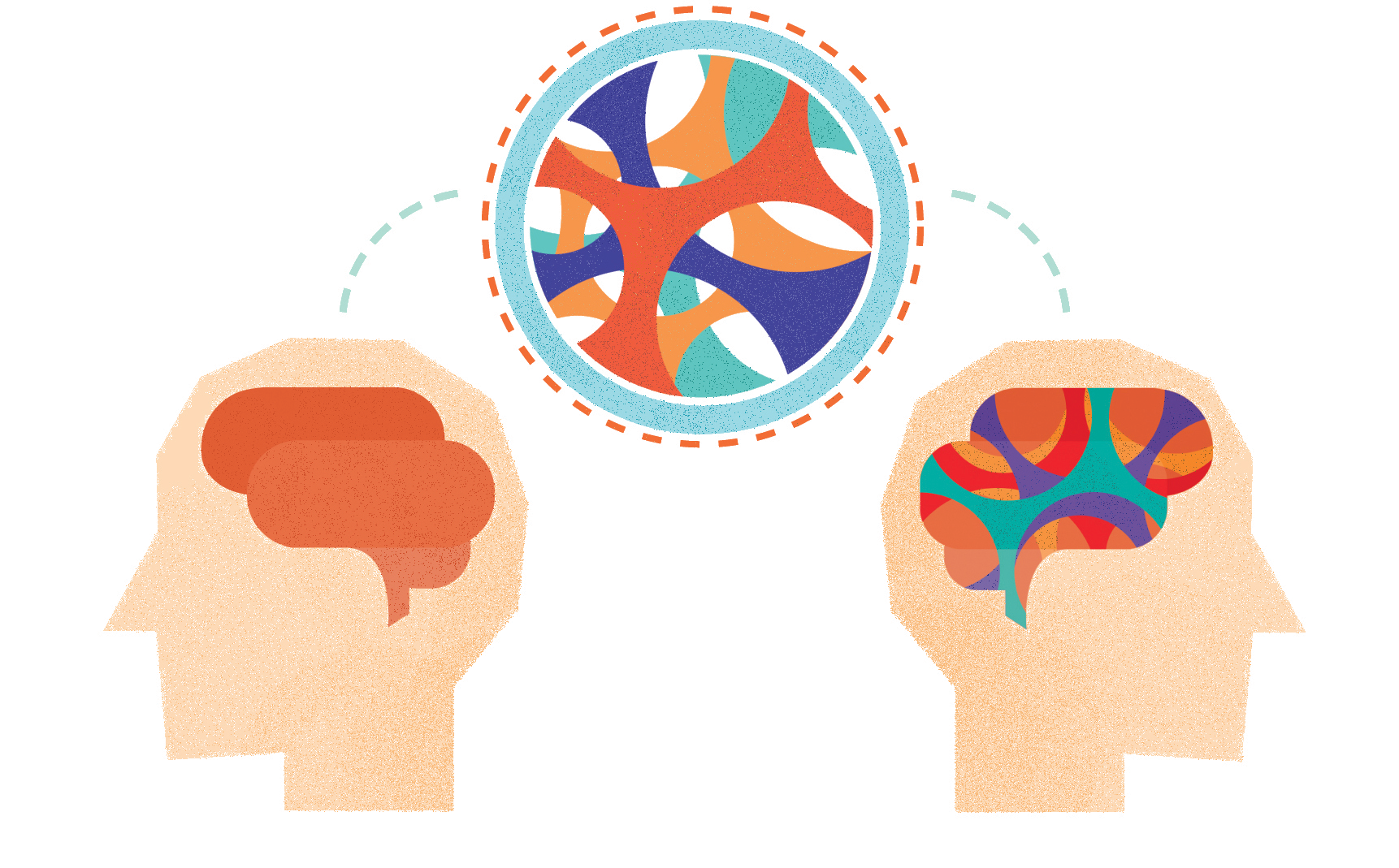
Historically, scientists believed the brain was “immune privileged,” meaning the immune system has little to no direct access to the central nervous system (CNS). The vessels necessary for immune cell circulation in the brain were first described in 1816; however, the scientific community discredited this discovery because it could not be reproduced [1][2]. It was not until 2015, almost 200 years later, that two groups independently rediscovered the existence of the anatomical structures allowing for proper immune function in the brain [3][4]. This discovery has radically changed the scientific community’s understanding of the relationship between the CNS and the immune system and has opened many new avenues for further research. The findings could potentially shed light on diseases that have been traditionally studied in the context of brain immunity.
The Immune Privileged Brain
The scientific community believed the brain was uniquely immune privileged as a result of the absence of lymphatic vessels, the vessels necessary for immune cell circulation, in combination with the selectivity of the blood-brain barrier [5, 6, 7]. Lymphatic vessels are just as numerous and extensive in the body as blood vessels, but circulate immune cells, called lymphocytes, rather than blood. Lymphocytes play an essential role in the body’s adaptive immune response by recognizing foreign pathogens and tagging them for destruction [5]. Although the circulatory system and lymphatic system are for the most part isolated from each other, open lymphatic vessels co-localize with areas that have a high density of capillaries, the thin blood vessels that allow for the diffusion of oxygen and nutrients into surrounding tissues [5]. Co-localization enables lymphocytes to interact with pathogens in the blood and mediate their destruction.
Before the discovery of lymphatic vessels in the brain, it was assumed that lymphocytes did not have access to blood circulating around the brain due to the selectivity of the blood-brain barrier. The blood-brain barrier functions as a division between the central nervous system and the rest of the body in order to protect the brain from potentially hazardous molecules that move through the bloodstream. Few molecules are small enough to pass the blood-brain barrier. While this effectively protects against many pathogens entering the CNS, it also prevents any antibodies from entering the CNS on the rare occasion that a pathogen manages to cross the blood-brain barrier, thus isolating the CNS from the full effect of an immune response [5][6].
Discovery of the Brain’s Immune Defense
Notably, although scientists thought the brain was largely sequestered from the immune system, a significant body of evidence suggested the existence of some immune functioning in the brain, carried out primarily by microglial cells [5][7]. Microglia are a class of glial cells that are responsible for a diverse range of functions in the brain, including the constant scanning of brain tissue in order to sense and respond to neurodegeneration, trauma, infection, and tumors in the brain. When microglia detect the presence of a pathogen, they may take on further diversified functions, such as effectively “eating” the pathogen or releasing chemical signals to alert other cells [5][7].
Although microglia were believed to be the primary mediators of immune function in the brain, it turns out there is more immune activity in the brain than previously thought. In June 2015, researchers at the University of Virginia studied the circulation of immune cells in dissected mouse brain meninges (the protective tissue that surrounds the brain) when they happened upon structured patterns of immune cell distribution [3]. The immune cells were highly concentrated in regions called the dural sinuses, which drain blood from vessels in the brain into the jugular veins. The team proceeded to assay for the presence of lymphatic endothelial cells and discovered vessels parallel to the dural sinuses as well as a network extending from the eyes to the olfactory bulb. Further study of the lymphatic vessels revealed they were capable of carrying immune cells in addition to draining cerebrospinal fluid (CSF) [3].
At the same time, researchers at the University of Helsinki reached similar conclusions about the existence of lymphatics in the brain [4]. These researchers studied lymphatics using a variety of modern imaging techniques and determined that “lymphatic-like” vessels exist in the eye, another organ traditionally believed to be immune privileged [8]. This discovery prompted further investigation, consequently leading to the independent discovery of lymphatics in the meninges of mice. These researchers observed extensive networks at the base of the skull, where the vessels exit the skull and transport CSF and macromolecules to deep cervical lymph nodes [4]. Preliminary findings suggest that similar structures exist in the human brain, but more data must be gathered before garnering a more substantial conclusion [3].

The Immune System and Behavior
Since the discovery of the brain’s immune system, further research into the interaction between the brain and immune system has revealed compelling connections between a class of signaling molecule used in the immune response, the interferon, and some aspects of our social behavior [9]. Interferon signaling plays an integral role in the immune response and is exhibited by both infected cells and immune cells. One particular interferon, IFN-γ, is expressed by meningeal lymphocytes. In experiments done on mice, rodents deficient in IFN-γ exhibited antisocial behaviors and the formation of excessive connections between neurons in the fronto-cortical regions of the brain, a phenotype implicated in autism spectrum disorder and schizophrenia [10][11]. Remarkably, when these mice were injected with IFN-γ, they lost all anti-social tendencies and showed a reduction of hyper-connectivity between neurons within twenty-four hours of administration [9].
This discovery and others like it may have great implications for researching and treating mental illnesses and neurodegenerative diseases associated with improper functioning of the immune system. For example, neurodegeneration was shown to trigger the recruitment of immune cells to the site of degeneration, leading to the formation of lesions characteristic of multiple sclerosis [12]. Another study found that immune cells interact with key proteins involved in Parkinson’s disease to promote neurodegeneration [13]. Deficiencies in proper immune functioning are also found in other neurodegenerative diseases, such as Alzheimer’s, and many mental disorders, including bipolar disorder, depression, schizophrenia, and alcohol addiction [14][15][16][17]. Although these discoveries by no means constitute a fast track to a cure, they certainly are an important step towards more clearly defining the limits of interactions between the brain and immune system.
Studies such as these may be very valuable in increasing the current body of knowledge regarding the relationship between our brains and immune systems in the context of disease states and mental disorders. The exact mechanisms behind many of these disorders are still poorly understood, but future research in this field may provide new hope in the form of novel prevention methods and treatments for improving mental health and well-being.
References
- Mascagni, P., and G.B. Bellini 1816. Istoria Completa Dei Vasi Linfatici. Vol. II. Presso Eusebio Pacini e Figlio, Florence. 195 pp.
- Lukić, I.K., V. Gluncić, G. Ivkić, M. Hubenstorf, and A. Marusić. 2003. Virtual dissection: a lesson from the 18th century. Lancet. 362:2110–2113. doi:10.1016/S0140-6736(03)15114-8
- Louveau, A., Smirnov, I., Keyes, T. J., Eccles, J. D., Rouhani, S. J., Peske, J. D., ... & Harris, T. H. (2015). Structural and functional features of central nervous system lymphatic vessels. Nature.
- Aspelund, A., et al., A dural lymphatic vascular system that drains brain interstitial fluid and macromolecules. J Exp Med, 2015. 212(7): p. 991-9
- Murphy, K., & Weaver, C. (2016). Janeway's immunobiology. Garland Science.
- Banks, W. A., & Erickson, M. A. (2010). The blood–brain barrier and immune function and dysfunction. Neurobiology of disease, 37(1), 26-32.
- Hanisch, U. K., & Kettenmann, H. (2007). Microglia: active sensor and versatile effector cells in the normal and pathologic brain. Nature neuroscience, 10(11), 1387-1394.
- Aspelund, A., Tammela, T., Antila, S., Nurmi, H., Leppänen, V. M., Zarkada, G., ... & Immonen, I. (2014). The Schlemm’s canal is a VEGF-C/VEGFR-3–responsive lymphatic-like vessel. The Journal of clinical investigation, 124(9), 3975-3986.
- Filiano, A. J., Xu, Y., Tustison, N. J., Marsh, R. L., Baker, W., Smirnov, I., ... & Peerzade, S. N. (2016). Unexpected role of interferon-γ in regulating neuronal connectivity and social behaviour. Nature, 535(7612), 425-429.
- Supekar, K., Uddin, L. Q., Khouzam, A., Phillips, J., Gaillard, W. D., Kenworthy, L. E., ... & Menon, V. (2013). Brain hyperconnectivity in children with autism and its links to social deficits. Cell reports, 5(3), 738-747.
- Whitfield-Gabrieli, S., Thermenos, H. W., Milanovic, S., Tsuang, M. T., Faraone, S. V., McCarley, R. W., ... & Wojcik, J. (2009). Hyperactivity and hyperconnectivity of the default network in schizophrenia and in first-degree relatives of persons with schizophrenia. Proceedings of the National Academy of Sciences, 106(4), 1279-1284.
- Scheld, M., Rüther, B. J., Große-Veldmann, R., Ohl, K., Tenbrock, K., Dreymüller, D., ... & Kipp, M. (2016). Neurodegeneration triggers peripheral immune cell recruitment into the forebrain. The Journal of Neuroscience, 36(4), 1410-1415.
- Pandey, M. K., Gross, C., Caballero, M., McKay, M. A., Tiwari, D., Schroeder, L. M., ... & Burrow, T. A. (2016). Immune cells encounter with α-synuclein fuels neurodegeneration in Parkinson’s disease. The Journal of Immunology, 196(1 Supplement), 51-5.
- Louveau, A., Da Mesquita, S., & Kipnis, J. (2016). Lymphatics in Neurological Disorders: A Neuro-Lympho-Vascular Component of Multiple Sclerosis and Alzheimer’s Disease?. Neuron, 91(5), 957-973.
- Bhattacharya, A., & Drevets, W. C. (2016). Role of Neuro-Immunological Factors in the Pathophysiology of Mood Disorders: Implications for Novel Therapeutics for Treatment Resistant Depression.
- Khandaker, G. M., & Dantzer, R. (2016). Is there a role for immune-to-brain communication in schizophrenia?. Psychopharmacology, 233(9), 1559-1573.
- Crews, F. T., & Vetreno, R. P. (2016). Mechanisms of neuroimmune gene induction in alcoholism. Psychopharmacology, 233(9), 1543-1557.
