For most people, the mere thought of a parasite setting up residence in their tissues is enough to induce a serious case of the creeps. There is something particularly horrifying about sharing something so personal (yeah, your insides) with an unwelcome intruder.
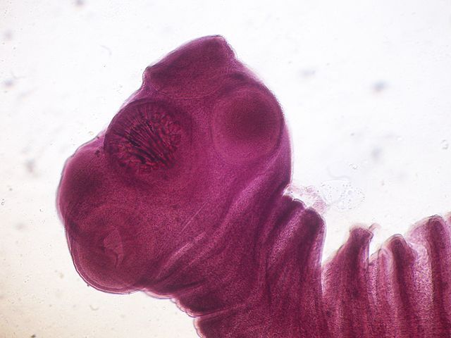
Taenia solium , better known as the pork tapeworm, is one such parasite. As an adult, the parasite attaches to the intestinal wall of its host (including humans) and grows anywhere from 2 to 7 m long producing an average of 50 million eggs 1 .
Figure 1. The life cycle of T. solum . Image source: the CDC .
However, more serious than the intestinal infection caused by the adult T. solum, is the infection caused by the larvae. The tapeworm is typically introduced into the human intestinal tract after consuming undercooked pork, where it then matures (Figure 1). But, if the eggs are consumed by another human (via inadvertent ingestion of contaminated fecal mater, for example) and develop into a larval stage within their host, the result is rather unsavory: larvae will burrow into the muscle and subcutaneous tissue of their host. This is an infection known as Cysticercosis 1,2 .
As unsettling as it may be to imagine these parasitic larvae burrowing into your muscles, it gets worse – both in clinical severity and general yuck-factor. A subset of cysticerosis infections is described by appending the prefix neuro .
Neurocysticerosis is the class of T. solium cyst infections of the central nervous system (CNS), which include the eyes, spinal cord, and brain 2 . Brain tissue is extremely sensitive to insult. As the tapeworm larvae burrow into brain structures, their occupancy disrupts brain function, which results in a series of clinically relevant neurological symptoms such as: migraine, psychiatric problems, arachnoiditis, vasculitis, and hydrocephalus 2,3 .
Among the most common symptoms is seizure 4,5 . Indeed, neurocysticercosis is the leading cause of epilepsy in underdeveloped communities and is becoming increasingly common in the United States 5 .
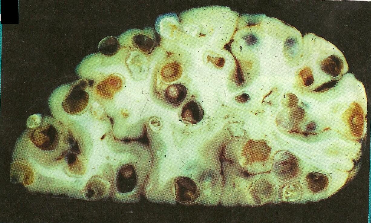
Figure 2 . In this brain many neurocysticercosis induced cysts have formed. Image source: Brane Space .
Efforts are being made to eradicate the prevalence of T. solium infections in endemic areas such as Latin America, Asia, and Africa. The World Health Organization is planning a pilot program in 2015, which, if successful, will be scaled “up in selected endemic countries by 2020. 6 ”
The pathology of all parasitic infections is interesting and neurocysticercosis is no exception. But, because it can infect the human brain, I find T. solium a bit more disgusting than the rest.
Interested in more parasites? Check out Carl Zimmer’s book on the subject .
References:
1. Centers for Disease Control and Prevention. http://www.cdc.gov/dpdx/taeniasis/index.html
2. Serpa, J. A., Machicado, J. (2013). Taenia solium and cysticercosis. ClinicalKey. Retrieved from: https://www-clinicalkey-com.offcampus.lib.washington.edu/#!/content/medical_topic/21-s2.0-2001693
3. Palacios, E., Lujambio, P.S., Jasso, R.R. 1997. Computed tomography and magnetic resonance imaging of neurocysticercosis. Seminars in Roentgenology, 32(4): 325-334.
4. Centers for Disease Control and Prevention. http://www.cdc.gov/parasites/cysticercosis/disease.html
DeGiorgio, C.M., Medina, M.T., Durón, R., Zee, C. Escueta, S.P. 2004. Neurocysticercosis. Epilepsy Currents. 4(3): 107-111.
5. The World Health Organization. http://www.who.int/mediacentre/factsheets/fs376/en/