
We have all experienced sleepless nights and know what it is like to suffer the consequences: inability to focus, decreased productivity, and feeling lethargic. Sleep is a process that is not fully understood. However, we do know that we spend a third of our life sleeping, so it must be important, right?
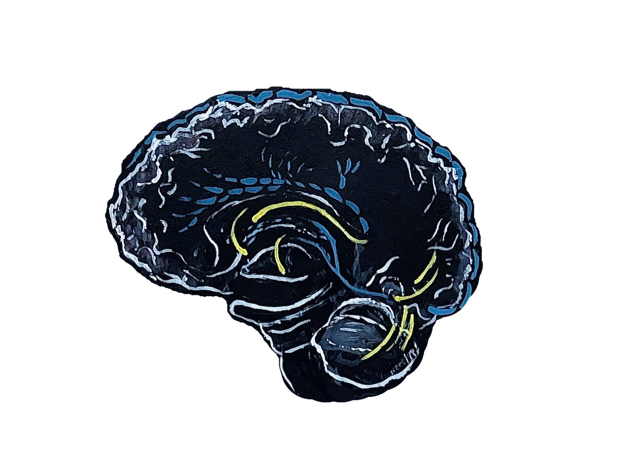
One of the reasons sleep is so important is because our bodies use that time to clear out unwanted byproducts, such as excess amino acids and nutrients [1]. Similar to other functioning systems, such as factories, our bodies produce said byproducts. Factories eliminate their byproducts by releasing them into the atmosphere. Similarly, humans eliminate many cellular waste byproducts via the lymphatic system, which is a network of highway-like vessels traveling throughout our bodies [2]. Until recently, it was thought that the same system runs through our brain and clears out the unwanted byproducts produced there. In the context of the brain, those materials are called solutes. However, in 2012, University of Washington Associate Professor Jeffrey Iliff discovered that a different system known as the glymphatic system performs this function in the brain [3]. This system is characterized by cerebrospinal fluid (CSF) flow in the brain, facilitating the movements of solutes out of the brain and into the lymphatic system, where they are subsequently removed [3]. The glymphatic system is mainly active during sleep and inhibited during wakefulness [1]. This suggests that for our brain to clear out its waste, we need sleep.
Mechanisms
Sleep is defined as a state of reduced mental and physical activity that generally occurs every night for around eight hours and is composed of multiple sleep cycles. A single sleep cycle is composed of two different stages of sleep: rapid eye movement (REM) sleep and non-REM sleep [4]. REM sleep is commonly known as the time of sleep when our brain is most active and when we typically experience our most vivid dreams. Non-REM sleep includes the stages following up to REM and is defined by the progression through deep sleep. The glymphatic system increases its activity during a part of non-REM sleep called slow-wave sleep [4]. This stage is characterized by the emission of delta waves from our brains, which are the slowest recorded brain waves in humans [5]. This suggests that slow-wave sleep is the most crucial type of sleep for the glymphatic system — and potentially for our brain’s health.
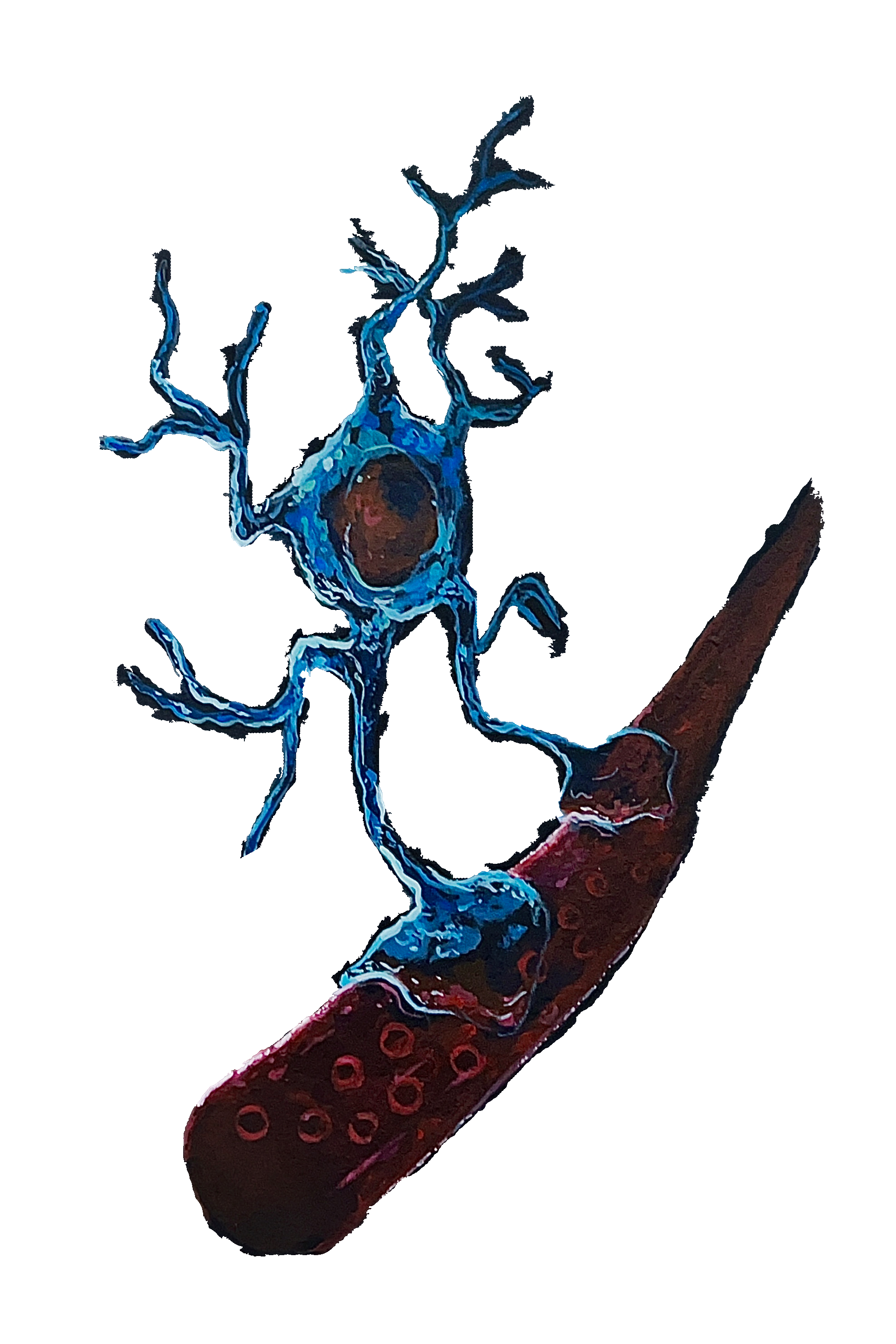
Our brain is populated by blood vessels, which supply nutrients to brain cells. Lining those blood vessels are highly specialized cells, called astrocytes, that are responsible for a great diversity of functions. Aquaporin 4 (AQP4) is a water channel that is found in those astrocytes [3]. During sleep, AQP4 channels localize at the endfeet of astrocytes, where they make contact with blood vessels [6]. This allows for the bulk flow of water out of the vessels and into the rest of the brain. Similarly to how a river flows and picks up sediment, the water picks up solutes as it moves through the brain and carries them away to where they can be cleared. When examining genetically modified mice that lack the AQP4 water channel, scientists observed a 70% decrease in the clearance of solutes compared to regular mice [7]. This supports the idea that AQP4 is a crucial contributor to glymphatic clearance.
Additionally, a recent study shows that the glymphatic system is not only sleep dependent but also circadian dependent [8]. The circadian rhythm is our body's 24-hour clock, and it has hormonal regulations that release time-dependent signals to our body. For example, it is the reason we wake up in the morning feeling awake and refreshed, and the reason we feel tired at night. When examining mice that were deliberately kept awake at their normal sleeping hours, it was found that although the animals were not in a sleep state, AQP4 channels still localized to the vessels. This led researchers to conclude that AQP4 not only localizes when you sleep, but also when you are supposed to be sleeping [8]. In other words, when an organism’s sleep pattern is disrupted short-term, it actually has little consequences on the efficacy of the glymphatic system. However, while the system certainly has components that are circadian regulated, the study shows that sleep-fragmented mice that are deliberately kept awake for consecutive nights show significantly increased solute deposition. This demonstrates that while sleep is not the only regulator of the glymphatic system, it is the most crucial. Thus, pulling an all-nighter now and then will not likely harm your brain long-term, but pulling multiple in a row is a recipe for disaster.
Lymphatic System Comparison
A similarly named system with a parallel purpose is called the lymphatic system– the waste clearance system for the rest of the body. It is characterized by a network of vessels that carry a fluid called lymph, and it functions by clearing cellular byproducts to lymph nodes which then transport the waste to our liver. Lymph nodes can be thought of as waste hotspots, in the same way that a garbage can localizes household waste to one place where it can be easily cleared. In addition to being responsible for waste removal, the lymphatic system is also responsible for the transportation of immune cells. Therefore, when we get sick, this system supports the allocation of immune resources to our area of infection or injury by assisting with the mobilization of T-cells, B-cells, and many other immune cells [9].
Being activated mostly at night is not the only distinguishing factor between the two systems. The glymphatic system is found strictly in our spine and brain, and utilizes vessels that are associated with blood as opposed to lymph in the lymphatic system. The glymphatic system vessels are lined by astrocytes as opposed to endothelial cells like in the lymphatic system. As previously discussed, astrocytes help maintain the flow of fluid between the brain and blood vessels using the localization of AQP4.
The main experiment that distinguished the two systems came in 2012 when Dr. Iliff and colleagues injected a contrast dye into the spine of mice [3]. In previous experiments where the injection took place outside of the brain and spinal cord in the mice, the dye spread to lymphatic vessels. However, in this case, live dynamic imaging showed that the dye stayed in the spinal cord and brain areas. This finding characterized the distinction between the lymphatic system and the glymphatic system [3].
Alzheimer's Disease
Recent studies have suggested that dysfunctions of the glymphatic system may be related to a variety of diseases, including Alzheimer's disease (AD) [10][11]. AD is a neurological disorder characterized by a decay in mental functioning, and it is currently the seventh leading cause of death in the United States. Experts estimate that one of every nine 65-year-olds and one of every six 80-year-olds living in the US are currently living with AD [12]. As the medical field continues to make advancements, the average life expectancy is only expected to increase, and with it, so will the number of people living with AD. Consequently, discovering more about the cause of AD and what we can do to prevent it is a crucial task.
One of the solutes the glymphatic system is responsible for clearing out is a protein called β-amyloid, a hallmark of Alzheimer’s disease (AD) [10]. This protein is created by billions of cells in our brain known as neurons. While the reasoning behind the formation of β-amyloid is not fully understood, we know that these proteins clump together between cells and disrupt their ability to communicate [10]. Researchers hypothesize that these aggregates also disrupt the flow of CSF through the brain, hindering its ability to clear out other solutes [11].
One of the most prominent characteristics of AD is the accumulation of β-amyloid plaques [11]. β-amyloid itself is a solute cleared by the glymphatic system, and so its disruption of the system leads to a positive feedback loop where increased β-amyloid aggregation leads to more blockage, which leads to more β-amyloid aggregation. This suggests that limiting the amount of β-amyloid aggregation by supporting glymphatic system function may be important in preventing AD. Since the glymphatic system is most active when asleep, a healthy sleep schedule is likely vital to brain health. One recent study shows that sleep-disrupted mice develop AD pathology significantly earlier compared to their sleep-control counterparts [11]. Furthermore, β-amyloid deposition as well as AQP4 mislocalization in the sleep-fragmented mice also increased, signifying the importance of sleep in the context of brain health [11].
Other factors that also are associated with the dysfunction of the glymphatic system by increasing the risk of AD are aging and head injuries. As we age, we experience a lower quality of sleep, caused by a range of factors, including less sleep time, frequent awakenings, and decreased slow-wave sleep [13]. As previously discussed, the glymphatic system has the best chance to clear the brain of solutes when sleep quality is high. Therefore, receiving inadequate sleep promotes the aggregation of β-amyloid and thus progresses the onset of AD. Additionally, traumatic brain injuries (TBIs) and other types of injuries to the head have been found to negatively affect glymphatic function [14]. When examining mice with and without a TBI, it was found that non-TBI mice show much greater clearance of solutes from the head [3]. Long-term effects of TBIs are well-known in humans. Experiencing a moderate TBI can increase your chance of suffering from AD by 2.3 times, while experiencing a severe TBI increases your chance by 4.5 times [14]. TBIs have an incredibly negative effect on brain health, especially in the context of glymphatic function and AD. Take football players for example: 14% of all National Football League players develop AD, and another 14% develop another form of dementia. This means that 28% of professional players end up developing a neurodegenerative disease, compared to the national rate of 10% in the general US population [15]. The increased rate of AD associated with TBIs further indicates the importance of sleep and glymphatic function.
The interplay of AD and glymphatic function can be modeled by a boxing fight. We are still unsure who threw the first punch and started the mess we know as Alzheimer's disease and glymphatic dysfunction, but we do know the characteristics of the fight. AD is caused by an increase of the deposition of β-amyloid in the brain, a protein that is partly cleared by the glymphatic system. The glymphatic system relies on sleep to function, primarily slow-wave sleep, and AD is associated with a decreased quality of sleep, increased sleep awakenings, and a decrease in overall sleep time, which includes slow-wave sleep. Additionally, it is hypothesized that the β-amyloid plaques that form in the brain cause a damming effect on glymphatic flow and thus decrease overall clearance, which in turn leads to more deposition. It seems that punches are being thrown back and forth and no one is winning– ultimately, both fighters lose together.
Conclusion
Many connections have been made between glymphatic function and dysfunction to various diseases and injuries. The most prominent ones include AD and TBIs. We see a clear pattern between the effect of TBIs on the onset of AD and the decreased efficacy of the glymphatic system. We also see that in severe cases of amyloid deposition, characteristic of AD, there is dysfunction of the glymphatic system. This ties together the importance of sleep to brain health.
The field of glymphatic system research exploded in 2012, when the system was recognized as an individual entity. Since then, the number of labs researching the glymphatic system has increased exponentially. Individual experiments are not able to isolate high-depth real-time glymphatic function yet due to technological deficiencies, but as technology advances, so will our understanding of the glymphatic system.
Similar to our other systems, the glymphatic system is a delicate one that could be negatively affected by a variety of factors. However, while technologies exist that can help us breathe or help our heart pump, there is nothing [yet] that can help clear our brain of β-amyloid and other solutes, preventing poor brain health, for example, the onset of AD. That is why sleep, a still largely unknown aspect of human life, needs to be one of our highest priorities when thinking about our health.
References
- Rasmussen, M. K., Mestre, H., & Nedergaard, M. (2018). The glymphatic pathway in neurological disorders. The Lancet. Neurology, 17(11), 1016–1024. doi.org/10.1016/S1474-4422(18)30318-1
- Swartz M. A. (2001). The physiology of the lymphatic system. Advanced drug delivery reviews, 50(1-2), 3–20. doi.org/10.1016/s0169-409x(01)00150-8
- Iliff, J. J., Wang, M., Liao, Y., Plogg, B. A., Peng, W., Gundersen, G. A., Benveniste, H., Vates, G. E., Deane, R., Goldman, S. A., Nagelhus, E. A., & Nedergaard, M. (2012). A paravascular pathway facilitates CSF flow through the brain parenchyma and the clearance of interstitial solutes, including amyloid β. Science translational medicine, 4(147), 147ra111. doi.org/10.1126/scitranslmed.3003748
- Scullin, M. K., & Gao, C. (2018). Dynamic Contributions of Slow Wave Sleep and REM Sleep to Cognitive Longevity. Current sleep medicine reports, 4(4), 284–293. doi.org/10.1007/s40675-018-0131-6
- Harmony T. (2013). The functional significance of delta oscillations in cognitive processing. Frontiers in integrative neuroscience, 7, 83. doi.org/10.3389/fnint.2013.00083
- Camassa, L. M. A., Lunde, L. K., Hoddevik, E. H., Stensland, M., Boldt, H. B., De Souza, G. A., Ottersen, O. P., & Amiry-Moghaddam, M. (2015). Mechanisms underlying AQP4 accumulation in astrocyte endfeet. Glia, 63(11), 2073–2091. doi.org/10.1002/glia.22878
- Mestre, H., Hablitz, L. M., Xavier, A. L., Feng, W., Zou, W., Pu, T., Monai, H., Murlidharan, G., Castellanos Rivera, R. M., Simon, M. J., Pike, M. M., Plá, V., Du, T., Kress, B. T., Wang, X., Plog, B. A., Thrane, A. S., Lundgaard, I., Abe, Y., Yasui, M., … Nedergaard, M. (2018). Aquaporin-4-dependent glymphatic solute transport in the rodent brain. eLife, 7, e40070. doi.org/10.7554/eLife.40070
- Hablitz, L. M., Plá, V., Giannetto, M., Vinitsky, H. S., Stæger, F. F., Metcalfe, T., Nguyen, R., Benrais, A., & Nedergaard, M. (2020). Circadian control of brain glymphatic and lymphatic fluid flow. Nature communications, 11(1), 4411. doi.org/10.1038/s41467-020-18115-2
- Breslin, J. W., Yang, Y., Scallan, J. P., Sweat, R. S., Adderley, S. P., & Murfee, W. L. (2018). Lymphatic Vessel Network Structure and Physiology. Comprehensive Physiology, 9(1), 207–299. doi.org/10.1002/cphy.c180015
- Meyer-Luehmann, M., Stalder, M., Herzig, M. C., Kaeser, S. A., Kohler, E., Pfeifer, M., Boncristiano, S., Mathews, P. M., Mercken, M., Abramowski, D., Staufenbiel, M., & Jucker, M. (2003). Extracellular amyloid formation and associated pathology in neural grafts. Nature neuroscience, 6(4), 370–377. doi.org/10.1038/nn1022
- Shokri-Kojori, E., Wang, G. J., Wiers, C. E., Demiral, S. B., Guo, M., Kim, S. W., Lindgren, E., Ramirez, V., Zehra, A., Freeman, C., Miller, G., Manza, P., Srivastava, T., De Santi, S., Tomasi, D., Benveniste, H., & Volkow, N. D. (2018). β-Amyloid accumulation in the human brain after one night of sleep deprivation. Proceedings of the National Academy of Sciences of the United States of America, 115(17), 4483–4488. doi.org/10.1073/pnas.1721694115
- (2023). 2023 Alzheimer's disease facts and figures. Alzheimer's and Dementia., 19: 1598-1695. doi.org/10.1002/alz.13016
- Li, J., Vitiello, M. V., & Gooneratne, N. S. (2018). Sleep in Normal Aging. Sleep medicine clinics, 13(1), 1–11. doi.org/10.1016/j.jsmc.2017.09.001
- (2023, May). Traumatic Brain Injury (TBI). Alzheimer’s Disease and Dementia, www.alz.org/alzheimers-dementia/what-is-dementia/related_conditions/traumatic-brain-injury#:~:text=Those%20with%20a%20history%20of,mild%20TBI%20increases%20dementia%20risk. Accessed 30 May 2023.
- Sauer, A. (2014, September 26). NFL Players Are at Increased Risk for Alzheimer’s. Alzheimers.Net, www.alzheimers.net/nfl-players-are-at-increased-risk-for-alzheimers.