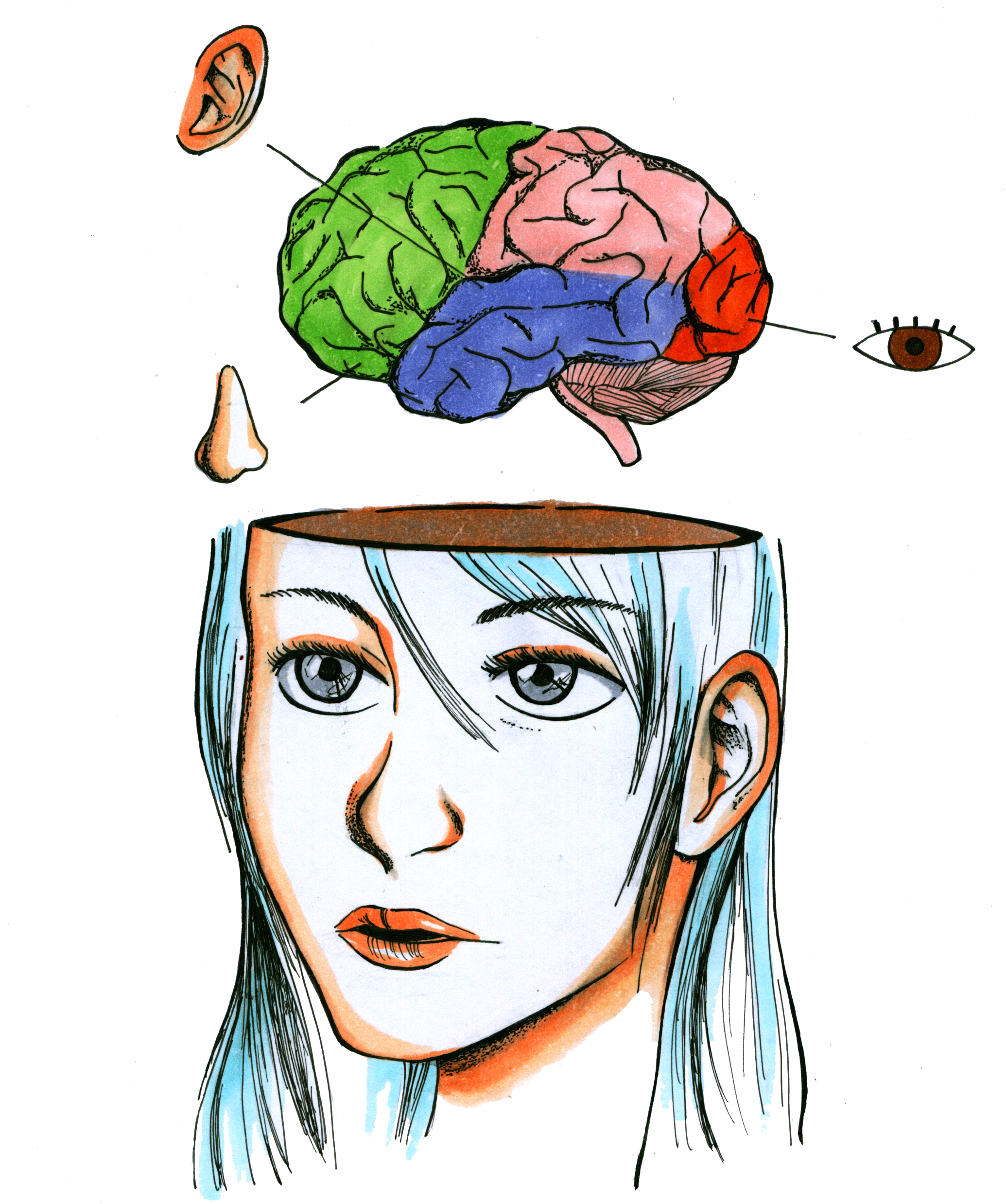
Our capacity to respond to, and communicate with, the outside world is contingent on the brain’s ability to both consciously and unconsciously process sensory information.
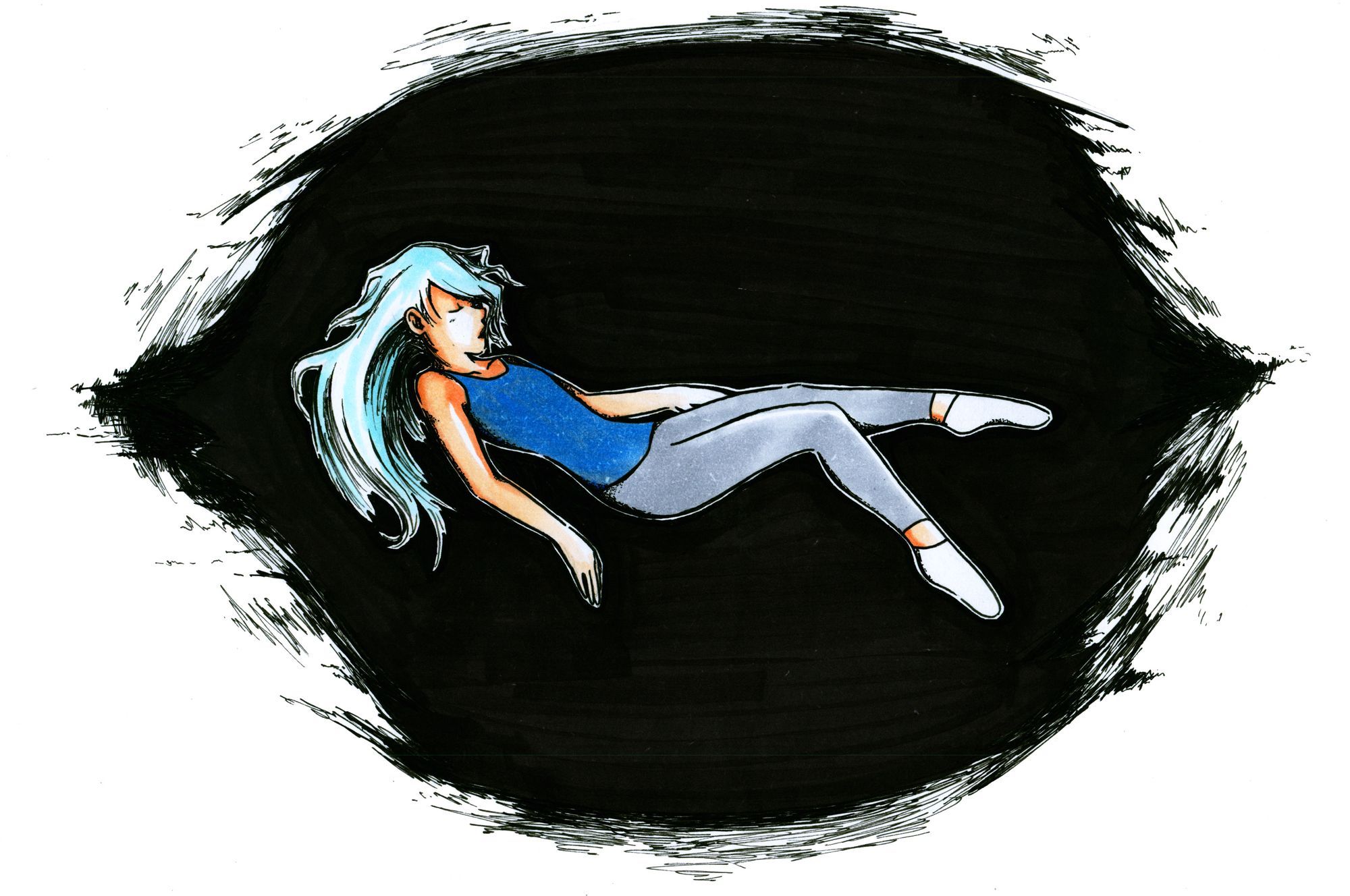
While sensation may seem like something that is consciously perceived, most sensory integration and processing occurs in the spinal cord, brainstem, and cerebellum without our conscious knowledge. Even during periods of sleep, the brain detects and responds to external stimuli. With such a vast network of neural circuitry devoted to the detection and processing of sensory information, it is interesting to imagine what might happen if the brain could be isolated from all sensory input. Interestingly, a new technique known as sensory deprivation therapy seems to simulate an environment almost completely devoid of sensory information. Although it may sound more like science fiction, sensory deprivation is now used in spas and is often referred to as “flotation therapy.” Subjects float in a pod filled with salt water that isolates them from sensory input such as light, sound, and touch. After this experience, users have reported experiencing intense hallucinations and feelings of regeneration. However, the mechanism behind how these chambers produce such extreme results remains unknown.
Restricted Environmental Stimulation Therapy
Also known as Restricted Environmental Stimulation Therapy (REST), sensory deprivation is generally offered in commercial spa locations. Subjects are placed in a chamber that is light-proof, soundproof, and scentless. It is partially filled with water containing magnesium sulfate, or Epsom salt, and heated to the temperature of human skin. The concentration of salt in the water is such that individuals float, and the temperature of the water makes it almost undetectable to the human body. Subjects feel a sense of zero gravity, and most sensory stimuli are absent [6].
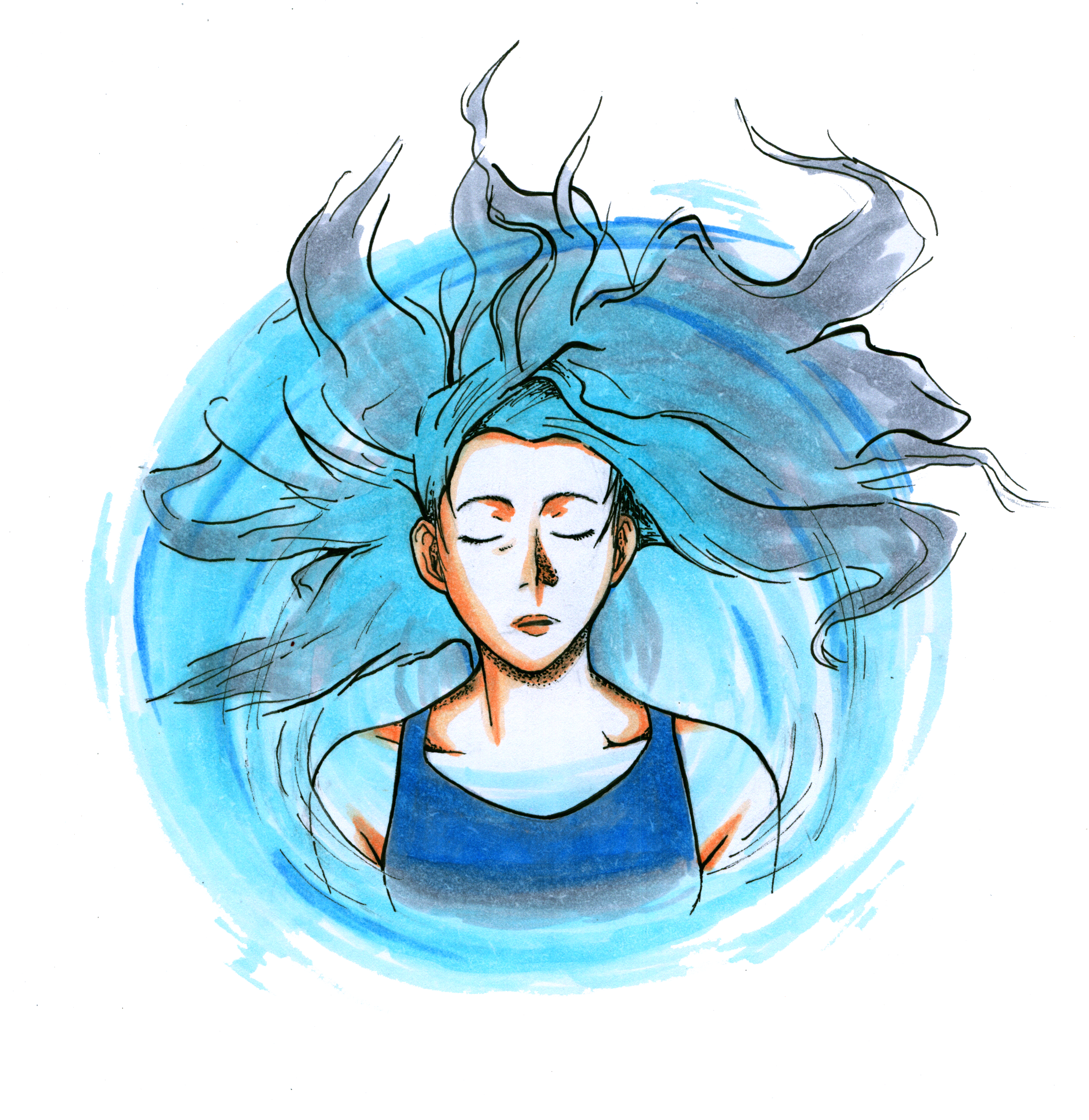
While REST attempts to eliminate many different types of sensory information, research studies and patient reports suggest that removal of even a single sense can elicit hallucinations [1]. Additionally, some subjects report that they experience the world in a more vibrant way following therapy, feeling more in tune with their own senses [6].
Removing One Sense at a Time
Although subjects often report heightened senses and visual hallucinations during and after their sensory deprivation experience, testing the therapy’s psychosomatic effects (effects that include the body and mind) is extremely difficult. The delicate equipment that is capable of detecting abnormalities in brain activity would need to be waterproofed and adapted to work in the isolation chamber. Additionally, to preserve the environment, the equipment could not add any sensory stimulation for the subject. Because of these difficulties, a combination of separate studies that test the effects of deprivation on a single sensory system can be used to gain some insight into how REST might affect the brain.
Light Deprivation
Scientists hypothesize that short-term light deprivation leads to increased excitability in the occipital cortex. This means that the occipital cortex is more sensitive and easily excited by sensory input. In order to test this hypothesis, researchers used Transcranial Magnetic Stimulation (TMS) to noninvasively stimulate this area of the brain [2]. TMS stimulation of the occipital cortex caused subjects to perceive flashes of light, known as phosphenes, in the absence of visual input. Researchers used this method to quantify changes in occipital cortex excitability by asking the subjects to report the appearance of phosphenes. Researchers then measured the “phosphene threshold,” the minimum intensity of TMS necessary to evoke phospehene perception. All subjects exhibited a decreased phosphene threshold during periods of light deprivation. The phosphene threshold returned to baseline levels within two hours of re-exposure to light [2]. Essentially, this means that when subjects experienced a period of light deprivation, it took significantly less stimulation to evoke these flashes of light. This suggests that the occipital cortex is more excitable during these periods of sensory deprivation. Thus, it is possible that internal neural activity that does not normally produce a visual perception may become an excitatory stimulus during light deprivation. This may explain the source of the visual hallucinations that can take place during periods of isolation from light, such as REST.
Some subjects also report seeing the world more vibrantly following sensory deprivation therapy. In order to better examine the potential cause of this phenomenon, researchers used functional Magnetic Resonance Imaging (fMRI) to measure neural activity in the occipital cortex during stimulation [2]. This technique was used in addition to TMS and does not rely on subject reports leading to more objective results than in the above-mentioned phosphene experiment. Experimenters found that subjects re-exposed to light thirty minutes following light deprivation displayed a significant increase in neural activity in the occipital cortex when compared to the control conditions, in which subjects were not exposed to light after deprivation [2]. This finding, which suggests an increase in the neural response to visual stimuli following subject re-exposure to light, may explain why subjects report seeing the world more vibrantly after sensory deprivation, as the brain may be more sensitive to visual stimuli.
Auditory Deprivation
Subjects also report experiencing auditory hallucinations during REST, as well as difficulty discriminating real and perceived sounds [6]. Researchers examined the potential mechanism behind this phenomenon by using a soundproof chamber to measure activity changes in auditory brain regions. Minneapolis, Minnesota is home to the quietest place on Earth, Orfield Lab’s anechoic chamber, with a sound level of -9 decibels [3]. For reference, the average sound level of a comfortable quiet space is about 30 decibels. All surfaces of the anechoic chamber absorb sound and are lined with soundproof fiberglass wedges that prevent sound reflection. People report hearing blood pumping in their heads in addition to their breathing, heartbeat, and digestive system. It is thought that the brain’s auditory systems enter a state of extreme activity due to the lack of external stimuli, and bodily sounds that are normally not heard become extremely loud [3]. To study the effects of short-term auditory deprivation, researchers recorded brainstem electrical responses of normally-hearing adults during and after auditory deprivation [4]. Subjects were exposed to auditory input after the deprivation period, which led to nerve fiber hyper-excitability, meaning that the brain became more sensitive to auditory input [4]. These findings may explain why subjects were unable to distinguish between real and perceived sounds, resulting in auditory hallucinations. Any stimulus created by the brain itself during a period of auditory deprivation may cause the brain to become hyper-excitable. The longest a person has lasted in the anechoic chamber is 45 minutes as these hallucinations often cause discomfort [3].
Smell Deprivation
Many subjects report experiencing senses in a more vivid way following REST, including their sense of smell. In order to test the effects of short-term smell deprivation, researchers conducted a study in which the subjects’ noses were plugged in order to see how the brain responds to deprivation of olfactory stimuli [5]. While they slept, subjects were allowed to breathe normally in a low-odor hospital room. In order for the brain to update information regarding odor, there needs to be a constant feed of olfactory input. Therefore, the brain tries to compensate for the lack of information so that the system does not break down [5]. Researchers found that following the smell deprivation, there was an increase in activity of the orbitofrontal cortex, a region associated with olfaction. Once unrestricted breathing was restored, the subjects actually experienced enhanced odor detection. These changes in the brain may explain why subjects report having a more sensitive sense of smell following sensory deprivation therapy.
In the Dark
Although the use of REST is largely commercialized, some research studies have suggested potential benefits on a more scientific level. Some studies have reported significant increases in scores on a standardized creativity test after REST [6]. Additionally, research has demonstrated that sensory deprivation improves performance in athletic and musical tasks [6]. It is difficult, however, to control all variables in sensory deprivation studies, leading to reduced certainty regarding their conclusions [7]. Although we cannot test the effects of removing multiple senses at once, combining the findings of multiple studies suggests that it is likely that sensory deprivation leads to hyper-excitability of the sensory regions of the brain. A recent increase of research in the field, however, will hopefully shed more light on this fascinating and ambiguous phenomenon.
References
- Mason, Oliver J., and Francesca Brady. "The Psychotomimetic Effects Of Short-Term Sensory Deprivation." The Journal of Nervous and Mental Disease: 783-85. Web. 4 Oct. 2015.
- Boroojerdi, B. "Enhanced Excitability of the Human Visual Cortex Induced by Short-term Light Deprivation." Cerebral Cortex (2000): 529-34. Print.
- Gardiner, Bryan. "Big Question: Why Can Silence Make You Hear Things That Aren’t There?" Wired.com. Conde Nast Digital, 19 May 2015. Web. 26 Oct. 2015.
- Decker, T.newell, and Susan W. Howe. "Short-term Auditory Deprivation: Effect on Brainstem Electrical Response." Hearing Research: 251-63. Print.
- "Brain Changes After a Stuffed Nose Protect the Sense of Smell." News Center. Northwestern University Feinberg School of Medicine, 13 Aug. 2012. Web. 20 Oct. 2015.
- Fan, Shelly. "Floating Away: The Science of Sensory Deprivation Therapy - The Crux." The Crux. 4 Apr. 2014. Web. 4 Oct. 2015.
- Fan, Shelly. "Interview with Dr. Peter Suedfeld: The Science of Sensory Deprivation Therapy." Neurorexia. 4 Apr. 2014. Web. 4 Oct. 2015.