Autism and epilepsy affect millions of people worldwide and have a profound impact on the lives of patients as well as their friends and family. These disorders are challenging to understand and treat, as patients vary widely in symptoms and causes, with dozens of genes implicated in both disorders. However, both disorders share some key symptoms, such as a relation to excitation levels in the brain and issues with sleep. Dravet Syndrome, a rare form of epilepsy which shares many symptoms with both autism and general epilepsy, is caused by a mutation in a single gene and can be reproduced in mice. Studying the DS mouse model can thus shed light on comorbid aspects of both autism and epilepsy, such as their impact on circadian rhythms and sleep.
Autism and epilepsy are relatively common disorders, with a 0.1% and 1% incidence, respectively [1][2]. The two disorders overlap in some areas, as both are hypothesized to be caused by an imbalance in the firing of inhibitory and excitatory neurons. Perhaps due to this connection, the two tend to co-occur, with 25% of autistic patients having some form of epilepsy and 15.2% of epileptics being diagnosed with autism. Additionally, patients with either or both of these conditions often experience problems with sleep, such as difficulties maintaining sleep at night, waking in the morning, or nocturnal seizures that interrupt sleep [3][4][5]. However, both disorders are linked to a broad spectrum of causes and symptoms, making them difficult to model, understand, and treat [1][2].
A rare type of epilepsy called Dravet Syndrome (DS) provides a convenient model that scientists can study to better understand aspects of epilepsy and autism in general, in addition to DS-specific changes [6]. DS appears during a child’s first year of life as febrile (heat-induced) seizures, which then morph into spontaneous seizures in adulthood. Patients with DS also experience arrested mental and physical development as well as behavioral disturbances [7]. Around 42% of patients develop photosensitivity, an exaggerated response to light, in their second year of life [6]. Scientists take interest in the disorder due to its simple causal mechanism, a mutation involving one gene: SCN1A, which codes for a part of a voltage-gated sodium channel, Nav1.1 [1][8][9].
Sodium channels let sodium ions, positively charged particles, flow into or out of neurons [8]. Nav1.1 gives entry into specific inhibitory neurons, which then allows the neurons to fire by changing their charge balance. Upon firing, inhibitory neurons release inhibitory neurotransmitters that prevent their target neurons from firing and contribute to the balance of inhibition and excitation in the brain. The SCN1A gene encodes the pore that sodium ions travel through in Nav1.1, making it a vital component of neuronal communication. Without it, inhibitory neurons fire substantially less than usual [8][10]. Decreased inhibition results in increased excitation, which can lead to a hyper-excitable, seizure-prone brain.

Approximately 70-80% of DS patients have mutations in the SCN1A gene, and there is evidence that the remaining 2030% may have mutations in other genes that affect SCN1A expression [11][6][12]. Mutations lead to changes in the protein the gene encodes. These changes can hamper protein function or even make it non-functional, which would then prevent inhibitory neurons from firing. By creating mice that lack the SCN1A gene in one strand of their DNA, scientists have established a direct relationship between SCN1A mutations and DS. If the SCN1A gene is altered so that the Nav1.1 protein cannot function, DS develops. In other words, a partial loss of function in Nav1.1 sodium channels is the cause of DS. The DS mouse model provides a means to understand some symptoms of autism and epilepsy through a related disorder. By selectively mutating SCN1A in the mouse brain, scientists can illuminate the function of the Nav1.1 sodium channel depending on brain region or cell type, which can give insights to symptoms such as sleep disturbances [11][6][12].
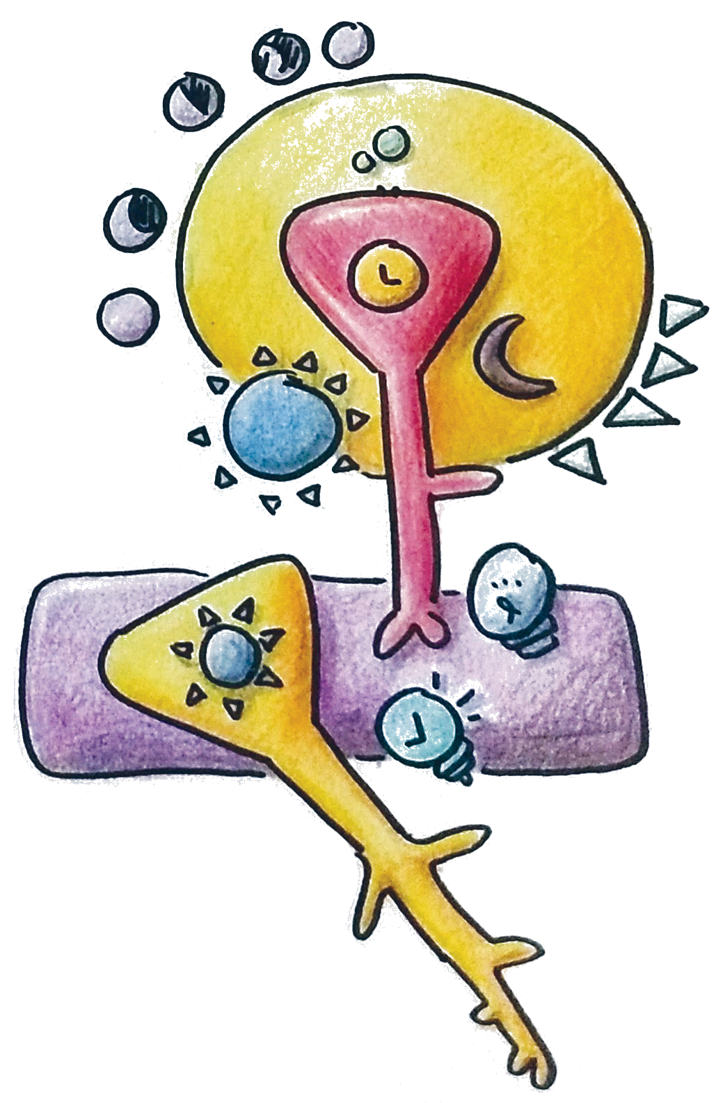
Patients with DS suffer from sleep problems much like patients with autism and epilepsy, reporting disturbances in sleep time and quality. Sleep-wake cycles are regulated by the suprachiasmatic nucleus (SCN), a grouping of cells within the hypothalamus that regulates a variety of circadian, or daily, rhythms [13]. For example, signaling from the hypothalamus changes levels of melatonin in the body, which then leads to the familiar cycle of sleeping at night and waking in the day. Other daily cycles include fluctuations in body temperature and hormone levels. Most neurons within the SCN are inhibitory and contain Nav1.1. Scientists propose that the loss of the Nav1.1 in DS mouse models impairs communication between the neurons in the SCN, thus reducing overall neuronal connectivity in the region [13].
Reduced connectivity in the SCN is implicated in sleep impairment [13]. The individual neurons in the SCN continue producing circadian rhythms on their own, as each neuron contains the molecular mechanisms necessary to produce periodic gene expression in a roughly 24-hour cycle. However, the neurons fail to coordinate with one another, so the overall output from the SCN does not create a regular fluctuation of signals that coordinate the daily rhythms of the body. Instead of producing the pronounced peaks and troughs of circadian gene expression, the SCN creates a somewhat level amount of expression [13][14].

A 50% loss of Nav1.1 also reduces signal transmission between the upper (dorsal) and lower (ventral) SCN [13][15][16]. The dorsal SCN receives information about the external light cycle and relays it to the ventral SCN, which takes action to adjust the biological clock accordingly. With less discrimination between day and night from the “master clock,” other biological clocks in the periphery, such as the liver circadian clocks that prepare for nutrient metabolism, can become uncoordinated as well [13][15][16][17][18].
Asynchrony leads to sleep-wake problems, as different clocks in the body may oppose one another on the true meaning of bedtime [15] . SCN1A mutant mice display less ability to discriminate between day and night, as they are less active than normal during their usual waking hours but more active than normal during their usual resting hours. Mutant mice also adapt poorly to changes in light rhythms, taking much longer to adjust [13]. In human terms, the mice are like people who get up frequently at night, feel lethargic during the day, and have a longer period of “jet lag” after moving through time zones. DS patients demonstrate similar symptoms, with difficulties entering and maintaining sleep. These traits are shared by around 4483% of autistic children, who are also reported to have a reduced difference between day and night in circadian metabolites like cortisol and melatonin [3][14]. In general epilepsy, sleep impairment is common among patients, with 40-55% of adults suffering from insomnia, 28-48% reporting daytime sleepiness, and more than 30% reporting that they sleep six hours or less [4].
However, sleep involves more areas of the brain than just the SCN, which primarily concerns its circadian aspect [18]. Sleep quality stems from the depth and integrity of sleep. DS mice and patients alike suffer in these regards, with patients often experiencing fragmentation of sleep, which hampers its integrity. Encephalographic studies demonstrate that DS mice also have reduced depth of sleep. These disturbances result from Nav1.1 loss in the reticular nucleus of the thalamus (RNT), a group of inhibitory neurons essential for the formation of structured, continuous sleep. They act together with the excitatory group of neurons in the ventrobasal nucleus, so reducing RNT function would create a mismatch between excitation and inhibition, causing sleep disturbances [18].
As seen both in the SCN and RNT, the DS mouse model supports the hypothesis that autism and epilepsy are caused by an imbalance of inhibition and excitation in the brain [19]. Further evidence for this theory comes from a study in which Nav1.1 was selectively deleted in DS mouse forebrain inhibitory neurons, excitatory neurons, or both. As expected, loss of Nav1.1 inhibitory neurons led to seizures; however, the same did not occur if the loss was in excitatory neurons. When both excitatory and inhibitory neurons lacked Nav1.1, the intensity of seizures was reduced, which indicates that reducing the imbalance between excitation and inhibition may lead to reduction of symptoms [19].
Although the DS mouse model provides a convenient means of studying DS, it is important to keep in mind that autism and epilepsy are still much more complex than DS; patients in both disorders vary greatly, both in terms of their genetics and their symptoms. Furthermore, not all of the therapeutic approaches that work in mice can “solve” the problems presented by the disorder, especially since DS is known to be “drug-resistant” [11].
However, DS models do indicate pharmacological interventions that improve symptoms for mice with partial Nav1.1 channel loss, which could apply to other symptoms specific to impaired inhibitory activity. A study found that clonazepam, a benzodiazepine, restored autism-like behaviors to normalcy in mutant mice by boosting the activity of the inhibitory neurons [20]. Another study, which ventured into modeling DS in zebrafish, found that clemizole can act as an anti-convulsant, and further proposed that modeling DS in zebrafish could be an effective way of testing drugs since the fish are easy to breed [21]. Although DS patients are already administered medication, drug testing and further investigation into the mechanisms of the disorder can reveal new methods with more specific strategies for alleviating symptoms.

Not all forms of autism and epilepsy arise from mutations in the Nav1.1 protein. However, by identifying the implication of Nav1.1 malfunction in sleep disorders and perhaps in other symptoms of both disorders, more specialized care can be provided. Drugs meant to target cells with Nav1.1 impairment could serve to reduce specific symptoms, perhaps administered with other treatments that target other specific mutations. Although DS is extremely rare, exploiting the Dravet Syndrome mouse model could lead to great discovery and advancements in the understanding of related disorders.
References
- Weiss, L. A., Escayg, A., Kearney, J. A., Trudeau, M., MacDonald, B. T., Mori, M., … Meisler, M. H. (2003). Sodium channels SCN1A, SCN2A and SCN3A in familial autism. Molecular Psychiatry , 8 (2), 186–194. https://doi.org/10.1038/sj.mp.4001241
- Stafstrom, C. E. (2014) Recognizing Seizures and Epilepsy, in Epilepsy (eds J. W. Miller and H. P. Goodkin), John Wiley & Sons, Oxford. doi: 10.1002/9781118456989.ch1
- Goldman, S. E., Alder, M. L., Burgess, H. J., Corbett, B. A., Hundley, R., Wofford, D., … Malow, B. A. (2017). Characterizing Sleep in Adolescents and Adults with Autism Spectrum Disorders. Journal of Autism and Developmental Disorders , 47 (6), 1682–1695. https://doi.org/10.1007/s10803-017-3089-1
- Jain, S. V., & Kothare, S. V. (2015). Sleep and Epilepsy. Seminars in Pediatric Neurology , 22 (2), 86–92. https://doi.org/10.1016/j.spen.2015.03.005
- Li, B.-M., Liu, X.-R., Yi, Y.-H., Deng, Y.-H., Su, T., Zou, X., & Liao, W.-P. (2011). Autism in Dravet syndrome: Prevalence, features, and relationship to the clinical characteristics of epilepsy and mental retardation. Epilepsy & Behavior , 21 (3), 291–295. https://doi.org/10.1016/j.yebeh.2011.04.060
- Wolff, M., Cassé-Perrot, C., & Dravet, C. (2006). Severe Myoclonic Epilepsy of Infants (Dravet Syndrome): Natural History and Neuropsychological Findings. Epilepsia , 47 , 45–48. https://doi.org/10.1111/j.1528-1167.2006.00688.x
- Harkin, L. A., McMahon, J. M., Iona, X., Dibbens, L., Pelekanos, J. T., Zuberi, S. M., … Scheffer, I. E. (2007). The spectrum of SCN1A-related infantile epileptic encephalopathies. Brain , 130 (3), 843–852. https://doi.org/10.1093/brain/awm002
- Meisler, M. H., & Kearney, J. A. (2005, August 1). Sodium channel mutations in epilepsy and other neurological disorders. https://doi.org/10.1172/JCI25466
- Nakayama, T., Ogiwara, I., Ito, K., Kaneda, M., Mazaki, E., Osaka, H., … Yamakawa, K. (2010). Deletions of SCN1A 5′ genomic region with promoter activity in Dravet syndrome. Human Mutation , 31 (7), 820–829. https://doi.org/10.1002/humu.21275
- Robbins, C. A., Kalume, F., Yu, F. H., McKnight, G. S., Burton, K. A., Mantegazza, M., … Spain, W. J. (2006). Reduced sodium current in GABAergic interneurons in a mouse model of severe myoclonic epilepsy in infancy. Nature Neuroscience, 9(9), 1142. https://doi.org/10.1038/nn1754
- Berkvens, J. J. L., Veugen, I., Veendrick-Meekes, M. J. B. M., Snoeijen-Schouwenaars, F. M., Schelhaas, H. J., Willemsen, M. H., … Aldenkamp, A. P. (2015). Autism and behavior in adult patients with Dravet syndrome (DS). Epilepsy & Behavior , 47 , 11–16. https://doi.org/10.1016/j.yebeh.2015.04.057
- Fujiwara, T., Sugawara, T., Mazaki‐Miyazaki, E., Takahashi, Y., Fukushima, K., Watanabe, M., … Inoue, Y. (2003). Mutations of sodium channel α subunit type 1 (SCN1A) in intractable childhood epilepsies with frequent generalized tonic–clonic seizures. Brain , 126 (3), 531–546. https://doi.org/10.1093/brain/awg053
- Han, S., Yu, F. H., Schwartz, M. D., Linton, J. D., Bosma, M. M., Hurley, J. B., … Iglesia, H. O. de la. (2012). NaV1.1 channels are critical for intercellular communication in the suprachiasmatic nucleus and for normal circadian rhythms. Proceedings of the National Academy of Sciences , 109 (6), E368–E377. https://doi.org/10.1073/pnas.1115729109
- Glickman, G. (2010). Circadian rhythms and sleep in children with autism. Neuroscience & Biobehavioral Reviews , 34 (5), 755–768. https://doi.org/10.1016/j.neubiorev.2009.11.017
- Kyeong, S., Choi, S.-H., Eun Shin, J., Lee, W. S., Yang, K. H., Chung, T.-S., & Kim, J.-J. (2017). Functional connectivity of the circadian clock and neural substrates of sleep-wake disturbance in delirium. Psychiatry Research: Neuroimaging , 264 (Supplement C), 10–12. https://doi.org/10.1016/j.pscychresns.2017.03.017
- Hastings, M. H., Reddy, A. B., & Maywood, E. S. (2003). A clockwork web: circadian timing in brain and periphery, in health and disease. Nature Reviews Neuroscience , 4 (8), nrn1177. https://doi.org/10.1038/nrn1177
- Hogenesch, J. B., Panda, S., Kay, S., & Takahashi, J. S. (2003). Circadian Transcriptional Output in the SCN and Liver of the Mouse. In D. J. C. Organizer & J. A. Goode (Eds.), Molecular Clocks and Light Signalling (pp. 171–183). John Wiley & Sons, Ltd. https://doi.org/10.1002/0470090839.ch13
- Kalume, F., Oakley, J. C., Westenbroek, R. E., Gile, J., de la Iglesia, H. O., Scheuer, T., & Catterall, W. A. (2015). Sleep Impairment and Reduced Interneuron Excitability in a Mouse Model of Dravet Syndrome. Neurobiology of Disease, 77, 141–154. https://doi.org/10.1016/j.nbd.2015.02.016
- Ogiwara, I., Iwasato, T., Miyamoto, H., Iwata, R., Yamagata, T., Mazaki, E., … Yamakawa, K. (2013). Nav1.1 haploinsufficiency in excitatory neurons ameliorates seizure-associated sudden death in a mouse model of Dravet syndrome. Human Molecular Genetics , 22 (23), 4784–4804. https://doi.org/10.1093/hmg/ddt331
- Han, S., Tai, C., Westenbroek, R. E., Yu, F. H., Cheah, C. S., Potter, G. B., … Catterall, W. A. (2012). Autistic-like behaviour in SCN1A+/- mice and rescue by enhanced GABA-mediated neurotransmission. Nature , 489 (7416), 385–390. https://doi.org/10.1038/nature11356
- Baraban, S. C., Dinday, M. T., & Hortopan, G. A. (2013). Drug screening in SCN1A zebrafish mutant identifies clemizole as a potential Dravet syndrome treatment. Nature Communications , 4 , ncomms3410. https://doi.org/10.1038/ncomms3410
