
Look at a fixed point. Now turn your head left, then right. What do you notice? Well, as your head moves in one direction, your eyes move the same number of degrees in the opposite direction to keep the image fixed in your field of view. This simple function, coordinated by the brain, is called the vestibulo-ocular reflex (VOR), and it is calibrated to head motion. A region of the brain involved in motor learning, called the cerebellum, is in part responsible for this adaptive calibration.
Neuroscientists use a variety of techniques such as optogenetics and behavioral methods to understand how the cerebellum regulates the VOR. These studies illuminate how the cerebellum learns as a whole, which can help us better understand the cerebellum during normal function and develop treatments for cerebellar impairments. These new techniques are revolutionizing the cerebellar field, and will exponentially increase progress toward these goals.
The Basics: Anatomy and Physiology of the Cerebellum
The cerebellum has long been considered essential for motor coordination. It contains 80% of the human brain’s neurons despite being only 10% of its mass [1]. The cerebellum’s best-known functions are balance and coordination, and the neural circuitry it uses to carry out these functions is remarkably standardized. The cerebellum’s sole output to the rest of the brain is from cells located in the cerebellar nuclei. These nuclei are surrounded by the cerebellar cortex, which provides the only input to the cerebellar nuclei nuclei via a special class of cells called Purkinje cells [2].
Purkinje cells receive roughly 200,000 inputs, the most of any cell in the entire nervous system [3]. Most of these inputs come from granule cells, the most common cell in the nervous system, outnumbering all other cell types combined. These make up the vast majority of cerebellar neurons as well, and they transmit their inputs to Purkinje cells via special axons called parallel fibers. Purkinje cells also receive another major input from climbing fibers. When a climbing fiber fires, the corresponding Purkinje cells produce a special type of long-lasting and multi-peaked action potential called a complex spike [2]. The firing of these two types of inputs to Purkinje cells determines the strength of Purkinje cell synapses. Specifically, during the VOR, eye movements may not perfectly cancel out head motion. In response to these mistakes, climbing fibers induce complex spikes in Purkinje cells, causing Purkinje cells to weaken synapses in parallel fibers whose firing produced the error, a process critical for learning [4].
Understanding the Cerebellum: Simple Behaviors
Reflexes like the VOR are examples of simple behaviors, and neuroscientists use them to study and identify the cerebellum’s less complex computations in order to gain information about higher-order processing that is more difficult to study. In simple cases, very few muscles are controlled to execute the movement. In contrast, an action such as reaching and grasping requires much more coordination, and thus exhibits neuron patterns which are more difficult to decipher. As our understanding of neural circuitry improves, we will be able to tackle more and more complicated behaviors.
Neuroscientists use behavioral methods to draw conclusions about cerebellar output and how it changes in response to various stimuli. Hold your hand up to be the fixed point your eyes watch, and observe your normal VOR by turning your head right, then left. Now, do the same head motion while moving your hand with your head as it turns, first in the same direction as head motion, and then in the opposite direction. When your hand moves with your head, there is essentially no VOR -- your eyes barely need to move to remain fixed on your head. However, when your hand moves in the opposite direction of your head, the VOR is amplified -- your eyes need to move more than normal to keep up. These changes in the VOR are a form of learning, and long-term change of this kind is a memory.
Studying how well the brain can learn to amplify or reduce the VOR under various conditions can help us understand what behaviors this neural circuit produces. We can then make better conclusions about how it works. For example, researchers used optogenetics, a technique in which flashing light near neurons causes them to fire through a genetically engineered chemical pathway, to identify the functions of certain neurons [5]. By manipulating the firing of climbing fibers during the VOR, they implanted a VOR memory in the brain that amplified the VOR. However, they were unable to stimulate climbing fibers in a way that reduced the VOR, showing that climbing fibers only drive learning during VOR amplification, not reduction. This work elucidated a new regulatory mechanism: climbing fibers regulate behaviors in the cerebellum, but the cerebellum may or may not respond to these error signals [5].
Cerebellar Learning: How We VOR
Error signaling provides a framework for a learning model of the cerebellum. Neurons learn through plasticity, which is a change in the connections between them. One type of plasticity is long-term potentiation (LTP), in which neurons that fire together experience a strengthening of connections and an increase in the number of synapses shared with each other. However, this does not apply to the majority of neurons in the brain. The cerebellum, which contains 80% of the brain’s neurons, primarily uses long-term depression (LTD) to learn, dropping the connections that produce errors instead of synthesizing new ones [4]. Given this principle, it is natural to expect that if we genetically engineered Purkinje cells to have enhanced machinery for LTD, the cerebellum would show enhanced learning of behaviors. However, for a particular kind of enhancement, this was not the case.
Scientists at Stanford University found that compared to normal mice, a particular transgenic mouse with enhanced machinery for LTD displayed impaired learning of VOR amplification [6]. They hypothesized that spontaneous, normal brain activity in the circuit induced LTD and dropped many neural connections before learning even occurred. As a result, during training, mice were unable to drop many connections to learn because there were few synapses still available to be dropped; LTD was completely saturated. This resulted in less VOR amplification in response to the same training compared to normal mice, in which very few connections were dropped prior to learning [6].
To test their hypothesis, the researchers designed an experiment to unsaturate the circuit and then train the brain to do VOR amplification directly after, so that the mice with enhanced LTD would learn faster than controls. They devised a pretraining protocol using VOR reduction beforehand to reduce the number of connections dropped before training began, leaving room for connections to be dropped later [6]. Their experiments showed data consistent with this hypothesis: the mice with enhanced LTD showed impaired learning under normal conditions, but enhanced learning when pretrained with VOR reduction [6].
In this case, researchers saw a perceived impairment become an enhancement under certain conditions, showing us that learning deficits may not be deficits under all circumstances. Not only does this provide us with new insight into how the cerebellum works, but it also shows us that individuals with what we call “impairments” may not always be impaired. Sometimes they may just be different; sometimes gifted in ways we do not understand.
Lights, Camera, Action: The Development of New Techniques
Current techniques are already showing us interesting things about how the cerebellum functions and providing insight into the true meaning of impairment. As research continues, newly developed techniques will help us gather more informative data so we can draw more accurate conclusions regarding the learning functions of the cerebellum. Two new techniques, Cal-Light and CaMPARI, aid neuroscientists in identifying populations of neurons that fire at certain times and in comparing the sizes and properties of these groups of neurons.
Both Cal-Light and CaMPARI are similar to optogenetics, wherein, as you may remember, light is used to stimulate firing in neurons. In these two techniques, neurons light up (fluoresce) if they fire while the light is on. With Cal-Light, neurons light up green in response to a light pulse. This provides researchers with information about which neurons are firing at which points in a particular behavior [7]. Researchers can then deactivate or stimulate those neurons to see what their specific roles are in the behavior [7]. With CaMPARI, all neurons of a certain cell type contain a green fluorescent protein, which irreversibly changes to red if they fire during a light pulse over a period of time [8]. This provides neuroscientists with an “activity snapshot” of the neuron population, not only identifying which neurons were active over a period of time, but also how many neurons were active compared with the entire population of cells [8].
More Than Motor Movements: The Cerebellum’s Expanding Role
Studying the cerebellum’s role in simple behaviors, such as the VOR, can help elucidate its underlying circuitry and computations. This information can enhance our understanding of the role of the cerebellum in more complex functions. While neuroscientists have often studied motor behavior to learn about the cerebellum, it is actually involved in much more. Scientists have long known of connections between the cerebellum and higher-order regions such as the prefrontal cortex and Broca’s area. Damage to the cerebellum has been shown to result in lower performance on general intelligence tests as well as tests of goal-orientated behaviors. However, as much as we know about the cerebellum’s role through these high-level measures, comparatively less is known about the circuits behind these functions. Part of the reason for this is that few animals have them, making them more difficult to find and study. Another reason is that because the cerebellum is so involved in motor tasks, it is hard to differentiate between the cerebellum’s motor commands and its higher-order commands during tasks that often must involve both.
Some recent research suggests that the cerebellum may be involved in reward pathways [9]. In this case, researchers genetically engineered neurons in mice to fluoresce when they fired in order to observe firing patterns during a reward task. They found that granule cells not only fired in the presence of a reward but also when the reward was expected, even if it was not delivered [9]. Other studies continue to explore the role of the cerebellum in non-motor functions and challenge the notion that the cerebellum’s sole purpose is motor coordination.
Acknowledging the cerebellum’s role beyond motor coordination means we also must consider its role in diseases and dysfunctions. The cerebellum is implicated in a wide range of disorders from dozens of different ataxias to autism spectrum disorders. Particularly, its role in autism has only become clear in the last two decades. Scientists pinpointed an abnormal connection between two human brain regions implicated in autism: one in the cerebellum, called right Crus I (RCrusI), and one in the cerebral cortex [10]. When they created a mutant mouse with this abnormal connection and inactivated RCrusI, the mice displayed social impairments and repetitive behaviors characteristic of autism. However, activating RCrusI alleviated these behavioral impairments. These results suggest that a cerebellar impairment is sufficient to produce autistic symptoms in mice, improving our understanding of autism and bringing us one step closer to a treatment [10].
Conclusion
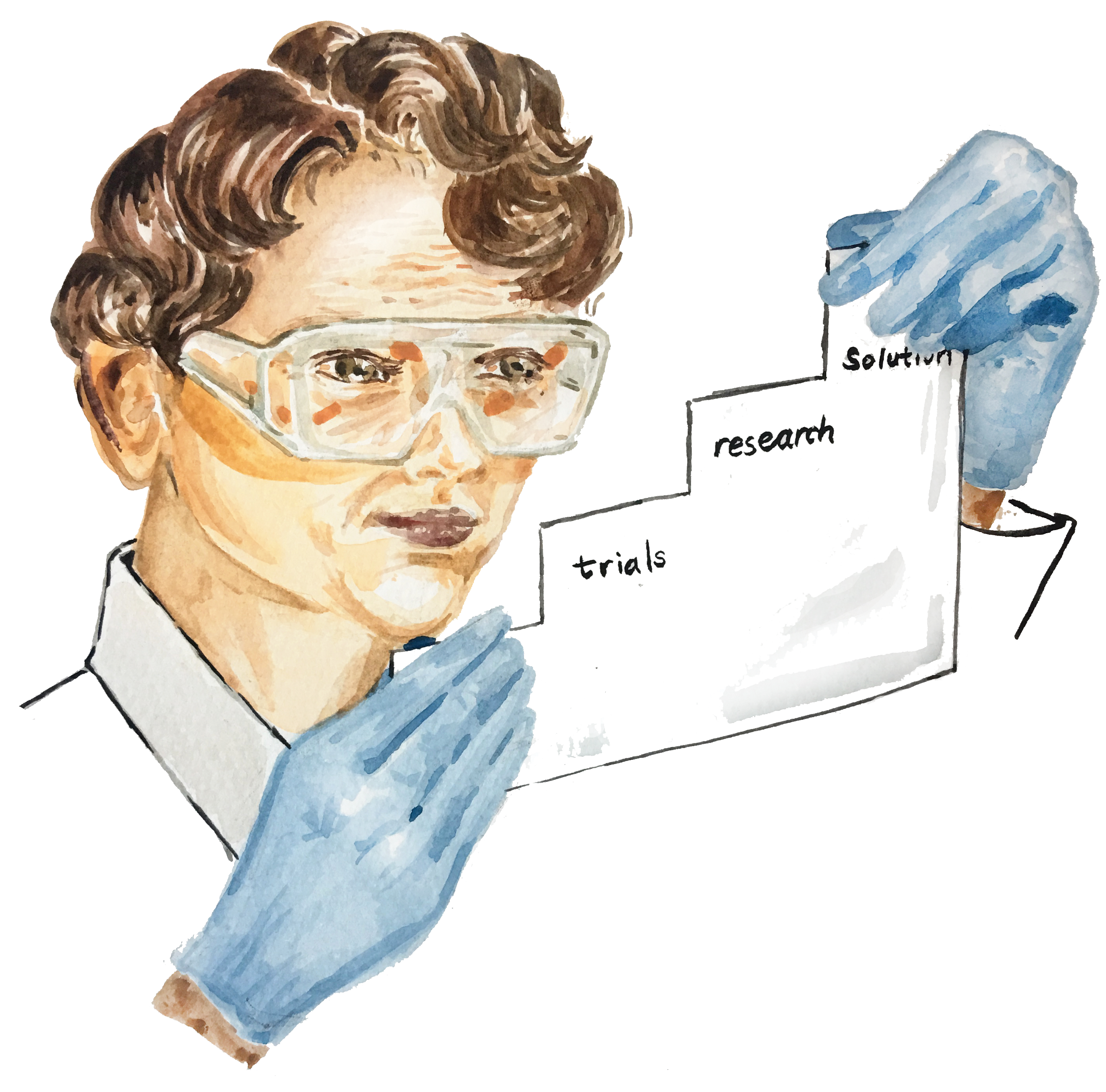
In order to reach big solutions, neuroscientists start small. Looking at a simple behavior in a well-understood brain region paves the way for understanding more complex behaviors and regions. Behavioral and optogenetic techniques help us understand the cerebellum so we can use that understanding to engineer solutions. Scientists who design and conduct these experiments are often thinking far into the future, beyond five or twenty or even eighty years, and are attempting to develop permanent solutions to some of the most widespread diseases afflicting humans today.
References
- Azevedo, F. A. C., Carvalho, L. R. B., Grinberg, L. T., Farfel, J. M., Ferretti, R. E. L., Leite, R. E. P., … Herculano-Houzel, S. (2009). Equal numbers of neuronal and nonneuronal cells make the human brain an isometrically scaled-up primate brain. The Journal of Comparative Neurology , 513(5), 532–541. doi:10.1002/cne.21974
- Raymond, J. L., Lisberger, S. G., & Mauk, M. D. (1996). The cerebellum: A neuronal learning machine? Science , 272(5265), 1126–1131. doi:10.1126/science.272.5265.1126
- Popa, L. S., Streng, M. L., & Ebner, T. J. (2018). Purkinje cell representations of behavior: Diary of a busy neuron. The Neuroscientist , 25(3), 241–257. doi: 10.1177/1073858418785628
- Grasselli, G., Hansel, C. (2014). Cerebellar long-term depression: cellular mechanisms and role in learning. International Review of Neurobiology, 117, 39-51. doi:10.1016/B978-0-12-420247-4.00003-8
- Kimpo, R. R., Rinaldi, J. M., Kim, C. K., Payne, H. L., & Raymond, J. L. (2014). Gating of neural error signals during motor learning. eLife , 3, e02076. doi:10.7554/elife.02076
- Nguyen-Vu, T. D. B., Zhao, G. Q., Lahiri, S., Kimpo, R. R., Lee, H., Ganguli, S., … Raymond, J. L. (2017). A saturation hypothesis to explain both enhanced and impaired learning with enhanced plasticity. eLife , 6, e20147. doi:10.7554/elife.20147
- Lee, D., Hyun, J. H., Jung, K., Hannan, P., & Kwon, H.-B. (2017). A calcium- and light-gated switch to induce gene expression in activated neurons. Nature Biotechnology, 35(9), 858–863. doi:10.1038/nbt.3902
- Fosque, B. F., Sun, Y., Dana, H., Yang, C. T., Ohyama, T., Tadross, M. R., … Schreiter, E. R. (2015). Labeling of active neural circuits in vivo with designed calcium integrators. Science , 347(6223), 755–760. doi:10.1126/science.1260922
- Wagner, M. J., Kim, T. H., Savall, J., Schnitzer, M. J., & Luo, L. (2017). Cerebellar granule cells encode the expectation of reward. Nature , 544(7648), 96–100. doi:10.1038/nature21726
- Stoodley, C. J., D’Mello, A. M., Ellegood, J., Jakkamsetti, V., Liu, P., Nebel, M. B., … Tsai, P. T. (2017). Altered cerebellar connectivity in autism and cerebellar-mediated rescue of autism-related behaviors in mice. Nature Neuroscience , 20, 1744-51. doi:10.1038/s41593-017-00