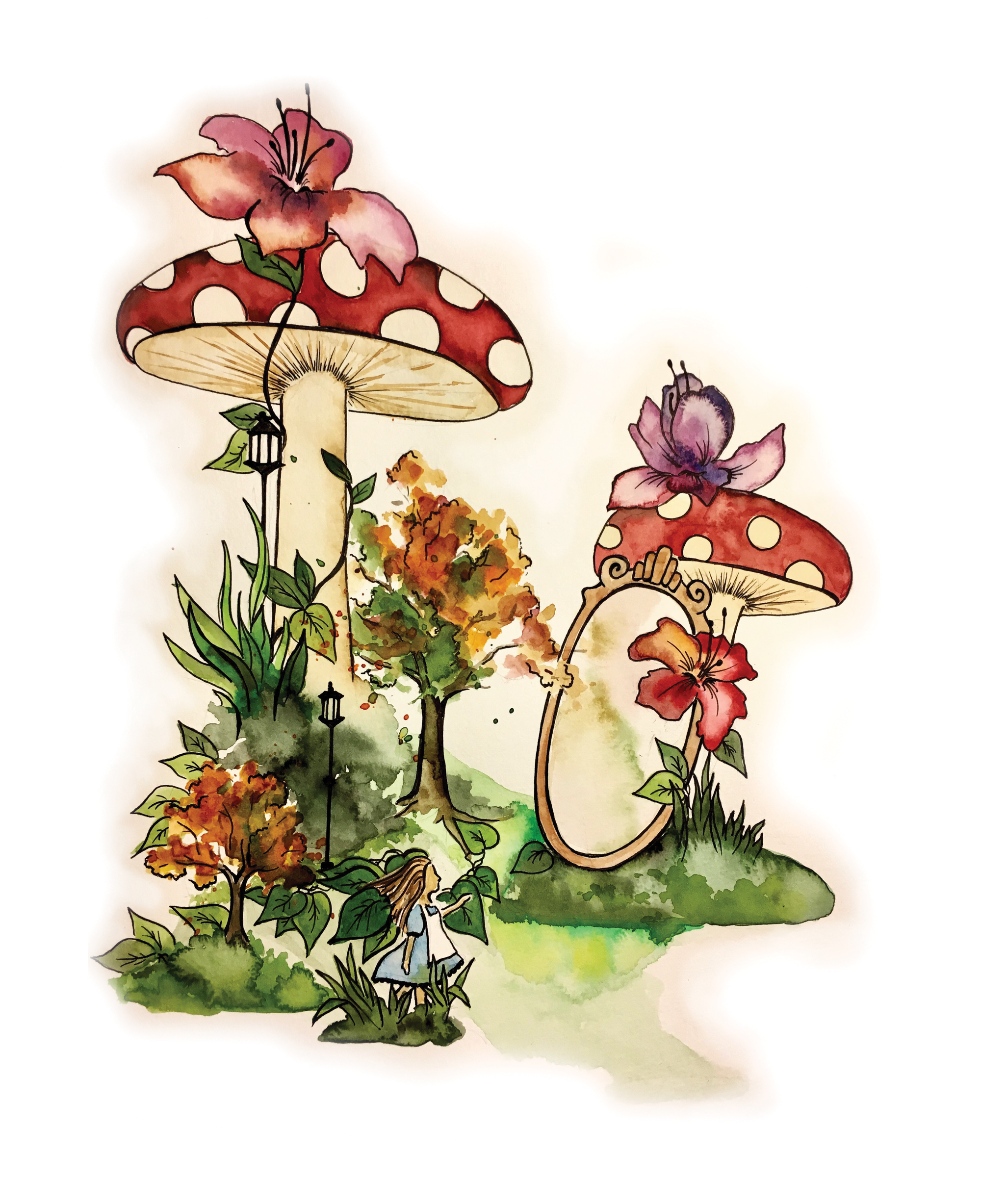
To some people, the world just doesn’t feel right. They may feel their bodies growing unnaturally large till they fill up the room. They sense that their bodies are shrinking to the size of a cup. These sudden distortions in the perception of body size are two of many disorienting symptoms experienced by those with a neurological condition known as the Alice-in-Wonderland syndrome (AIWS) . Identified by English psychiatrist John Todd in 1955, he named the syndrome after the protagonist in “Alice’s Adventures in Wonderland” (1865) by Lewis Carroll [1]. In Carroll’s original story, Alice followed a talking white rabbit down a rabbit hole and then experienced several dramatic changes in body size and shape. Her body first shrunk down to ten inches after drinking from a bottle she found, and shortly thereafter had a dessert that made her grow so large that her feet became impossible to see. Besides body image distortions, AIWS is also characterized by visual illusions, auditory hallucinations, impaired sense of time [2]. Patients can feel disconnected from the world, or observe themselves from outside their bodies, and might even have a feeling of being separated into two halves of their bodies. Over the past 60 years, AIWS symptoms have come to include 42 visual symptoms and 16 symptoms from other senses [3]. For example, the patient may experience macropsia, a condition where the individual sees everything larger than it actually is, micropsia, which means perceiving things to be smaller than it actually is, or telopsia, a condition where objects are perceived to be further than they actually are.
The syndrome is generally considered rare because fewer than 170 cases of AIWS have been documented or diagnosed [4]. However, clinical studies among patients with migraine indicate that 15% of the group experience AIWS symptoms such as micropsia, macropsia, telopsia [3]. The inconsistency is due to the fleeting passage of AIWS and the lack of major medical issues associated with the perceptual peculiarities [5]. Since most episodes are short and harmless, people who experience it would be unlikely to report it to a medical professional [3]. Therefore, it is difficult to ascertain with certainty whether Alice in Wonderland syndrome affects more people than the numbers estimated in the medical literature.

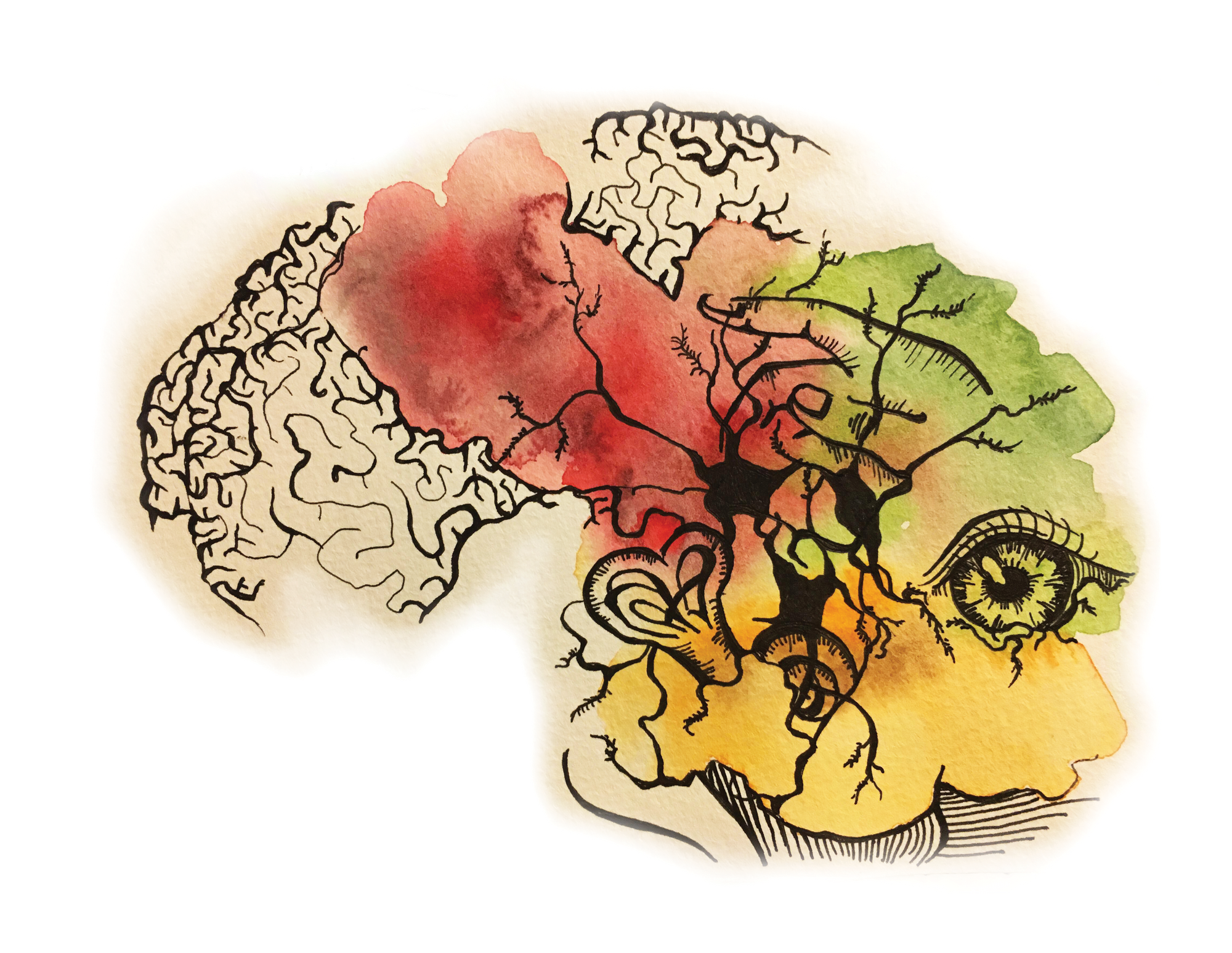
Underlying Neurobiology
AIWS can be caused by a variety of factors such as epilepsy, migraines, and infectious diseases [1]. It can also be induced by psychotic disorders or drug use. However, a link with migraines is suggested by the high comorbidity, or the presence of both diseases at once. Migraines are the most common cause of AIWS in adults (27.6%) and the second in children (26.8%). Interestingly, several child patients experiencing AIWS originating from causes other than migraine had a relevant family history of migraines and eventually developed migraine headaches themselves. This result is confirmed by a study on young students with visual illusions: one-third of subjects with episodic illusions were probable migraineurs [6]. Todd suggested a parietal location for the distortions of body image, and recent reports of patients with AIWS have shown dysfunction of the temporo-occipital or the parieto-occipital cortex – two brain regions heavily involved in information processing.
The association between migraine and AIWS may be explained by the neuronal effect of depolarization wave of neurons, called cortical spreading depression (CSD), on the aforementioned brain areas [8]. AIWS is more frequent in children and therefore may depend on structural differences between children’s and adult’s brain: associative brain areas are the last to develop and myelinate. Thus, the immaturity of associative areas, the temporo-occipital and parieto-occipital cortices, could explain how these are more vulnerable to spreading depression than in adults equally affected by migraine. To date, the close association between migraine and AIWS supports this idea, but at present this remains a speculation [4].
Studies also suggested that the symptoms of AIWS are attributed to functional and structural aberrations of the perceptual system [3]. Most symptoms of AIWS are attributed to centrally located neuron populations and even cell columns that respond selectively to specific types of sensory input. Area V4 of extrastriate visual cortex, for example, plays a role in color processing, whereas area V5 neurons encode the direction an object is moving. Both areas also respond to shape and depth, but bilateral loss of function of V4 results in the inability to see color, and bilateral loss of V5 results in the inability to see motion. Similarly, various neuron populations have been identified as being responsible for mediating different types of metamorphopsia, seeing straight lines curvy or rounded [3]. Sometimes AIWS symptoms involves aberrations between larger components of the visual network, which can vary individually. The same would seem to hold true for sensory distortions, in the sense that functional or structural anomalies of specific neuron populations in sensory areas are responsible for mediating body schema illusions. Whether similar mechanisms are responsible for mediating time distortions is still unknown. [3]
Another cortical area associated with AIWS is TPO-C, which is the crossroad of temporooccipital, parietooccipital, and temporoparietal junctions [4]. TPO-C is where visual and sensory information are integrated. AIWS may depend on either a direct functional change in the TPO-C or an impairment of the primary visual sensory cortices, which leads to the over-activation of areas responsible for spatial attention tasks. The spectrum of symptoms appears to be wider and more complex if abnormalities involve more anterior regions of the brain. In fact, theoretically, if an abnormality is located in occipital regions, we are more likely to observe simple visual disturbances. When the abnormality is closer to parietal and temporal areas, somatosensory and cognitive disorders will combine with the visual disturbances ones, resulting in more complex symptoms, as the areas integrate more abnormalities [4].
The complicated nature and the absence of a univocally accepted diagnostic criteria makes it quite difficult to analyze the various causes. The exact pathways of the syndrome depend on the underlying disorder each individual AIWS cases are associated with.
Treatment
Because the pathways leading to AIWS are still ambiguous, AIWS has no standardized treatment plan. Specific treatment is directed at underlying conditions each case is linked with. Thus, treatment requires a careful assessment using blood tests, EEG and brain MRI scans, as well as a better understanding of the progression of AIWS symptoms [3]. Depending on the individual’s particular disorder, doctors may prescribe antiepileptics, migraine prevention medication, antiviral agents, or antibiotics. Fortunately, most AIWS symptoms themselves are harmless and patients are typically discharged quickly after proper treatment.
Conclusion
Despite the findings, AIWS is still poorly known and probably misdiagnosed due to the lack of clear and universally accepted diagnostic criteria. After 60 years of relative obscurity, AIWS has begun to receive scientific attention again. This renewed interest is in part because of the growing possibilities to explore the brain’s networks responsible for mediating its symptoms with the aid of rapidly developing imaging techniques [3]. The underlying pathways are of great complexity. Although only a minority of AIWS cases reported specific information on site of brain correlate, it appears consistent that brain alterations responsible for AIWS are located in TPO-C, where the dorsal and ventral streams of visual system are integrated with somatosensory and vestibular inputs [4]. Better anatomical and functional studies of AIWS underlying mechanisms are needed, but a main prerequisite is a clear and univocally accepted classification system based on precise diagnostic criteria. Although AIWS is still a mystery, it provides an opportunity for us to study and understand the sensory perception in visual and sensory processes of the nervous system. With advancement in neuroimaging techniques, scientists should be able to further explore the mystery and understand our perceptual system better.
References
- Lanska, D. J., & Lanska, J. R. (2017). The Alice-in-Wonderland Syndrome. Neurologic-Psychiatric Syndromes in Focus Part II – From Psychiatry to Neurology Frontiers of Neurology and Neuroscience, 142–150. doi: 10.1159/000475722
- Jia, Y., & Miao, Y. (2018). Evidence for the Perception of Time Distortion During Episodes of Alice in Wonderland Syndrome. The Journal of Nervous and Mental Disease, 206(6), 473–475. doi: 10.1097/nmd.0000000000000825
- Blom JD. Alice in Wonderland syndrome: a systematic review. Neurol Clin Pract. (2016) 6:259–70. 10.1212/CPJ.0000000000000251
- Mastria, G., Mancini, V., Viganò, A., & Piero, V. D. (2016). Alice in Wonderland Syndrome: A Clinical and Pathophysiological Review. BioMed Research International, 2016, 1–10. doi: 10.1155/2016/8243145
- Naarden, T., Meulen, B. C. T., Weele, S. I. V. D., & Blom, J. D. (2019). Alice in Wonderland Syndrome as a Presenting Manifestation of Creutzfeldt-Jakob Disease. Frontiers in Neurology, 10. doi: 10.3389/fneur.2019.00473
- Abe K., Oda N., Araki R., Igata M. Macropsia, micropsia, and episodic illusions in Japanese adolescents. Journal of the American Academy of Child & Adolescent Psychiatry. 1989;28(4):493–496. doi: 10.1097/00004583-198907000-00004.
- Todd J. The syndrome of Alice in Wonderland. Canadian Medical Association Journal. 1955;73(9):701–704.
- Fine E. J. The Alice in wonderland syndrome. Progress in Brain Research. 2013;206:143–156. doi: 10.1016/b978-0-444-63364-4.00025-9.