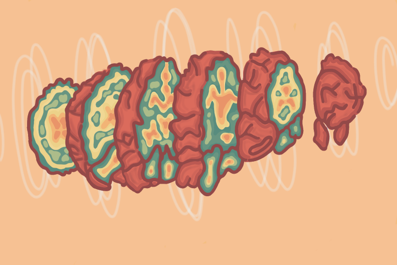
Have you ever thought about going to a professional when something about your body did not feel right? “Jerry,” a 73-year-old man, was in the same situation. He suffered from persistent memory problems and decided to see a neurologist. The neurologist listened to Jerry’s symptoms and without hesitation, diagnosed him with Alzheimer’s Disease (AD). However, over time it became obvious to Jerry’s family his symptomatology was not consistent with Alzheimer’s and they advised him to seek a second opinion. Jerry’s new doctor suggested that Jerry receive a Single Photon Emission Computed Tomography (SPECT) scan of his brain. Jerry and his family had never heard about SPECT scan imaging but decided to give it a try. When the scan was performed, it displayed a distinct lobster pattern indicative of enlarged ventricles, a characteristic sign of NPH.The SPECT scan also showed no signs of neuronal atrophy in the frontal lobe of the brain, a common sign of Alzheimer’s [1]. With the help of the SPECT scan, Jerry was correctly diagnosed with normal pressure hydrocephalus (NPH), an abnormal buildup of cerebrospinal fluid (CSF) in the brain's ventricles [2]. After the implantation of a thin tube to drain excess CSF away from the brain, Jerry’s memory markedly improved. Without the SPECT scan, the diagnosis and treatment of Jerry’s problem would not have been possible. However, our story does not end here. Jerry’s questions may have been answered, but ours haven’t. What is SPECT and how is it used? Why was it effective and why couldn’t specialists use a different brain imaging technology? SPECT has been around for more than twenty years [3]. Despite its longevity, it is not being used to its full potential due to a poor understanding of the scan. However, when used correctly, it can help optimize the treatment for complex cases, such as cognitive decline, NPH, OCD, ADHD, and PTSD [3].
What is required for SPECT to work?
SPECT is a nuclear imaging technique that produces a three-dimensional image of the patient’s blood flow. This helps provide detailed physiological information about the tissues in a targeted area [3]. Before a scan, patients must first undergo testing to see if they meet the necessary criteria for a SPECT scan. Patients who are pregnant or have a high caffeine intake are at high risk and should not get a SPECT scan [4]. After the patient meets the necessary qualifications, they are injected with a tracer compound. The tracer compound consists of a detectable radioactive isotope bound to a biologically active ligand, in other words, a molecule that binds to receiving protein transmitting a signal into the cell of the specific tissue being imaged [3][5]. Although radiation is involved in the SPECT scan process, the average radiation exposure of the SPECT imaging is 0.7 Roentgen equivalent man (rem).This is equivalent to the average annual exposure dose to aircrew who fly 800 hours per year [6]. When a tracer compound is injected into the patient, healthy tissues take up a known amount of the radioactive isotope and appear as a bright area in the SPECT image. Conversely, damaged/diseased tissues take up less or more of the radioactive isotope and show up as dark or more intense spots, respectively [7]. Technetium-99m (Tc) is the most commonly used isotope by clinicians. It is considered the most practical because of its short half-life of six hours. Shorter half-lives of an isotope lead to shorter scan times and less radiation exposure to the target organ. Also, compared to the other isotopes it has a higher sensitivity and specificity of the image. Another benefit of Tc is as the dose increases, the image quality also increases [8]. However, these mechanisms do not describe a SPECT scan’s advantages over other imaging techniques mentioned in the introduction. Let’s compare each common imaging technique and a SPECT scan’s advantages over each one to understand why SPECT is such an important tool.
SPECT equipment
The equipment used for SPECT imaging has to have 360 degrees rotation of the camera, otherwise, if we use 180 degrees rotation, it would lead to incomplete data and result in distortion in the reconstructed images [9]. Consequently, reconstructed images are affected by the image resolution, which has to be no less than a 128 × 128 image matrix meaning the camera will take 128 slices and put them on top of each other creating a picture. Following the previous sentence,fewer slices will lead to a lower resolution, and lower resolution will result in unclear images, where the smaller parts of the imaging will not be distinguishable [9].
SPECT scanning uses a multi-headed gamma camera that rotates around the patient during the scan and detects photons produced by the gamma decay of the tracer compound [10]. These cameras capture the emitted photons and convert them to light and then to a voltage signal, which is reconstructed into a final, observable planar image [11]. The detector is rotated around the patient, taking pictures every 3 to 6 degrees, which combine to make a 3D image [12].
Both the American College of Radiology [13] and the European Society of Nuclear Medicine (ESNM) [14] have published similar evidence-based guidelines for using SPECT scans to enhance diagnosis. These indications include:
- Evaluating patients for cerebrovascular disease
- Evaluating patients with suspected dementia
- Localizing seizures arising from a specific part of the brain
- Evaluation of traumatic brain injury
- Diagnosing encephalitis, inflammation of the brain, often due to infection
- Assessing brain death
Why is SPECT better than PET/fMRI?
Two compounds, Tc isotope and a compound known as hexamethylpropyleneamine oxime (HMPAO), are often used together for SPECT brain imaging [15]. HMPAO acts as the blood-brain barrier(BBB) transfer compound. The BBB serves to protect neural tissue from infectious diseases traveling within the bloodstream and is selective with what compounds it allows to cross. HMPAO is one of these special compounds able to transfer across. Therefore HMPAO is recognized as a metabolic substance by the brain tissue. These scans are able to show a patient’s brain activity and metabolically damaged tissues that would not be seen in the other types of tomography images [15]. For instance, functional magnetic resonance imaging (fMRI) scans show the current neurological activity of the brain reacting to a specific stimulus and the structure of the brain. On the other hand, SPECT is not as influenced by what is happening at the moment and shows a long-term pattern of brain function. SPECT scan differs from Positron emission tomography (PET) scans in that it only image areas where blood flows because its tracer remains in the bloodstream rather than being absorbed by surrounding tissues like in PET scan [16]. Also, the test has shown that SPECT scan is more sensitive to brain injuries than both MRI and PET scans because it shows reduced blood flow in injured areas.The ability to show function and brain activity by imaging where and how the tracer compound interacts with brain tissue makes SPECT scans especially useful in determining complex psychiatric cases which will be discussed later on in the paper.

Pros/Cons
However, SPECT scans also have notable disadvantages. SPECT images achieve their high-quality image over the 30-minute period of canning, during which the camera will rotate around the patient while they are lying flat on their back. Consequently, SPECT scans do not record an instantaneous change in brain activity like a fMRI. The whole procedure from the injection of the isotope to acquiring the image usually takes 1-2 hours. Compared to 45 minutes for an fMRI, SPECT scans do take slightly longer. However, compared to the PET scans which duration averages to 2 hours, SPECT scan is more beneficial. It is also important to note that in rare cases, patients may experience an allergic reaction to the radioactive tracer or ligand. Lastly, SPECT scans have a poor soft tissue contrast, making it challenging to point to the exact location of the affected brain tissue, what it shows is the area/ lobe that was affected [1]. However, when used with a CT scan the soft tissue contrast significantly improves leading to precise determination of the disease [17].
Although there are some downsides, SPECT scans have many advantages. One such advantage is the use of high-resolution cameras that allow for higher-quality images. While many may think this requires longer scan times, these higher-quality images can be achieved with shorter scan times and less radiation exposure compared to other imaging technologies [18]. SPECT imaging also provides visual images corresponding to the anatomy and functions of the brain.
Case study
SPECT imaging is a useful tool in psychiatry that has the potential to help millions of misdiagnosed people around the world [19]. A prime example of SPECT’s necessity in the medical community is illustrated in a case study regarding a man named Chase [19]. As a teenager, he was diagnosed with ADHD and bipolar disorder, which is usually described as a cycle between depression and mania [3]. Chase struggled throughout college and his career with severe anxiety, mood swings, crippling panic attacks, disrupted sleep and depressed mood. After years of jumping from one medication to the other with no improvement, Chase’s stepmother, who had been to a SPECT clinic in New York, recommended Chase go in for an evaluation.
When Chase arrived, doctors took a detailed history, performed neuropsychological tests, ran a lab workup, and performed a SPECT scan of his brain to see his brain blood flow and functional patterns. As the SPECT scan showed Chase’s brain had multiple areas with reduced amounts of blood flow that are commonly associated with dysfunctions related to memory, executive function, social interaction and impulse control [20]. Also, his scan showed multiple areas with increased perfusion of blood in the areas that are often associated with anxiety and depression. The combination of these patterns is consistent with the patients who were diagnosed with bipolar disorder [20].
Chase’s scan showed significantly low blood flow to his prefrontal cortex and temporal lobes. The prefrontal cortex is a region responsible for focus, judgment and impulse control, and the temporal lobes regulate mood stability, memory, and temper control [19].These results showed he had past head trauma and toxicity exposure, but not bipolar disorder. As it turns out, Chase used to race at a NASCAR speedway, where he suffered a number of significant concussions and was exposed to toxic gasoline fumes. Misdiagnosis of bipolar disorder after a significant concussion is common due to the similarity of symptoms and affected brain areas. SPECT imaging is a reliable way to distinguish these two conditions.
To treat Chase, doctors replaced his medications with brain-supportive supplements such as vitaminD and iron, common supplements for managing the effects of concussions. After eight months of treatment, his brain began to show significant improvements, and Chase had a considerable improvement in quality of life. Chase was given incorrect diagnoses based on his list of symptom clusters, and received medication that promised to improve their quality of life. However, a SPECT scan would have promptly indicated another course of action. Often, without actually looking at the brain, it is almost impossible to accurately find and treat the problem. Without a SPECT scan, Chase could have suffered throughout his whole life and might have never received a correct diagnosis. In addition to Chase’s case the SPECT has been used in other 1000 patients to diagnose PTSD, depression and bipolar disorder.
Future research/directions
With the current evolution of the gamma cameras and therefore improvement of the image quality of SPECT scans, in the future SPECT will be able to achieve better performance and allow the technology to be more widely used in clinical psychiatry to correctly diagnose patients [21]. The current barriers that stay in the way of SPECT technology are the sensitivity and spatial resolution compared to the other scanning methods. However, as the research shows SPECT is a rapidly changing field with new developments in technology and image-processing algorithms produced in the past several years, which in the future have a high chance of solving the sensitivity and spatial resolution problems [22]. Also, as mentioned in the Pros/Cons section, new technology introduces and encourages a diagnostic-quality hybrid SPECT/CT systems which are proven to have improved anatomic localization and diagnostic certainty. The quality and accuracy of SPECT imaging has been improved with technological progress and the improved gamma cameras reduced SPECT’s difference between predicted and actual diagnosis to as low as 12% [23]. It is exciting to see what the future holds for this technology and how it will be used in the medical field to improve brain disorder diagnosis.
Conclusion
Despite SPECT's rise in popularity psychiatrists remain skeptical of its validity due to the widely held idea that SPECT is restricted by limited resolution. However, as mentioned above the new improved multi-headed cameras produce resolution images and are consistently improving without being inferior to other scans. Furthermore, psychiatry is an evolving field, and if used correctly SPECT has the potential to become more popular and recognized as an important tool for clinicians to identify a patient's brain pathophysiology. With the use of SPECT brain imaging, clinicians will be able to examine the biological mechanisms behind the potential patient’s pathophysiology. A greater understanding of their unknown illness may lead to more relevant considerations about the patient’s past injuries, traumas, and experiences, and thus a better treatment plan and diagnosis. Utilizing neuroimaging tools such as SPECT will move psychiatry forward and help normalize brain health and medical treatment for psychological disorders.
References
- Dr. Allen, M. P. (2022, May 23). FMRI vs. SPECT Scan for the Brain: Know Your Options. https://www.cognitivefxusa.com/blog/fmri-vs-spect-scan-for-brain
- Normal pressure hydrocephalus. (2019, November 19). https://www.hopkinsmedicine.org/health/conditions-and-diseases/hydrocephalus/normal-pressure-hydrocephalus
- Hutton BF. The origins of SPECT and SPECT/CT. Eur J Nucl Med Mol Imaging. 2014 May;41 Suppl 1:S3-16.
- Committee Opinion No. 723: Guidelines for Diagnostic Imaging During Pregnancy and Lactation. Obstet Gynecol. 2017 Oct;130(4):e210-e216.
- What are Ligands? (2020, October 9). News-Medical.Net. https://www.news-medical.net/life-sciences/Ligands-An-Overview.aspx
- Health effects of ionising radiation on people. (n.d.). Retrieved November 30, 2022, from https://www.nea.gov.sg/our-services/radiation-safety/understanding-radiation/health-effects-of-ionising-radiation-on-people
- Alenizi , D., Kizilbash, N. A., Gill, O., Abukanna, A., Malik, S., & Badawy, A. (2013). Correlation of SPECT imaging, biochemical parameters and mutation with systolic dysfunction.
- Kane, S. M., & Davis, D. D. (2022). Technetium-99m. In StatPearls. StatPearls Publishing. http://www.ncbi.nlm.nih.gov/books/NBK559013/
- Unterrainer, M., Eze, C., Ilhan, H., Marschner, S., Roengvoraphoj, O., Schmidt-Hegemann, N. S., Walter, F., Kunz, W. G., Rosenschöld, P. M. af, Jeraj, R., Albert, N. L., Grosu, A. L., Niyazi, M., Bartenstein, P., & Belka, C. (2020). Recent advances of PET imaging in clinical radiation oncology. Radiation Oncology, 15(1), 88. https://doi.org/10.1186/s13014-020-01519-1
- Murphy, A. (n.d.). Digital image | radiology reference article | radiopaedia. Org. Radiopaedia. https://doi.org/10.53347/rID-53396
- Yandrapalli, S., & Puckett, Y. (2022). Spect imaging. In StatPearls. StatPearls Publishing. http://www.ncbi.nlm.nih.gov/books/NBK564426/
- Ha, S., Hong, S. H., Paeng, J. C., Lee, D. Y., Cheon, G. J., Arya, A., Chung, J.-K., Lee, D. S., & Kang, K. W. (2015). Comparison of spect/ct and mri in diagnosing symptomatic lesions in ankle and foot pain patients: Diagnostic performance and relation to lesion type. PLoS ONE, 10(2), e0117583. https://doi.org/10.1371/journal.pone.0117583
- Juni JE, Waxman AD, Devous MD, Tikofsky RS, Ichise M, Van Heertum RL, Carretta RF, Chen CC., Society for Nuclear Medicine. Procedure guideline for brain perfusion SPECT using (99m)Tc radiopharmaceuticals 3.0. J Nucl Med Technol. 2009 Sep;37(3):191-5.
- Graham M Guidelines and Standards Committee, Comments Reconciliation Committee. American College of Radiology Practice Guideline for the Performance of Single Photon Emission Computed Tomography (SPECT) Brain Perfusion and Brain Death Studies.
- Gawne, P. J., Man, F., Blower, P. J., & T. M. de Rosales, R. (2022). Direct cell radiolabeling for in vivo cell tracking with pet and spect imaging. Chemical Reviews, 122(11), 10266–10318. https://doi.org/10.1021/acs.chemrev.1c00767
- Noori-Asl, M. (2020). Investigation of different factors affecting the quality of spect images: A simulation study. Journal of Medical Physics, 45(1), 44. https://doi.org/10.4103/jmp.JMP_88_19
- Kapucu OL, Nobili F, Varrone A, et al. EANM procedure guideline for brain perfusion SPECT using (99m)Tc- labeled radiopharmaceuticals, Version 2. Eur J Nucl Med Mol Imaging. 2009;36:2093–102.
- Koppula BR, Morton KA, Al-Dulaimi R, Fine GC, Damme NM, Brown RKJ. SPECT/CT in the Evaluation of Suspected Skeletal Pathology. Tomography. 2021 Oct 11;7(4):581-605. doi: 10.3390/tomography7040050. PMID: 34698290; PMCID: PMC8544734.
- Khalil MM, Tremoleda JL, Bayomy TB, Gsell W. Molecular SPECT Imaging: An Overview. Int J Mol Imaging. 2011;2011:796025.
- Best SRD, Haustrup N, Pavel DG. Brain SPECT as an Imaging Biomarker for Evaluating Effects of Novel Treatments in Psychiatry-A Case Series. Front Psychiatry. 2022 Jan 13;12:713141. doi: 10.3389/fpsyt.2021.713141. PMID: 35095582; PMCID: PMC8793864.
- Amen, D. G. (2020). The end of mental illness: How neuroscience is transforming psychiatry and helping prevent or reverse mood and anxiety disorders, ADHD, addictions, PTSD, psychosis, personality disorders, and more. Tyndale Momentum, the nonfiction imprint of Tyndale House Publishers.
- Madsen, Mark T. “Recent Advances in SPECT Imaging.” Journal of Nuclear Medicine, vol. 48, no. 4, Apr. 2007, pp. 661–73. jnm.snmjournals.org, https://doi.org/10.2967/jnumed.106.032680.
- Jansena, Floris P., and Jean-Luc Vanderheyden. “The Future of SPECT in a Time of Pet - Researchgate.net.” The Future of SPECT in a Time of PET, https://www.researchgate.net/profile/Jean-Luc-Vanderheyden/publication/5924075_The_future_of_SPECT_in_a_time_of_PET/links/5dfa54cba6fdcc2837291134/The-future-of-SPECT-in-a-time-of-PET.pdf?origin=publication_detail.
