I. Introduction
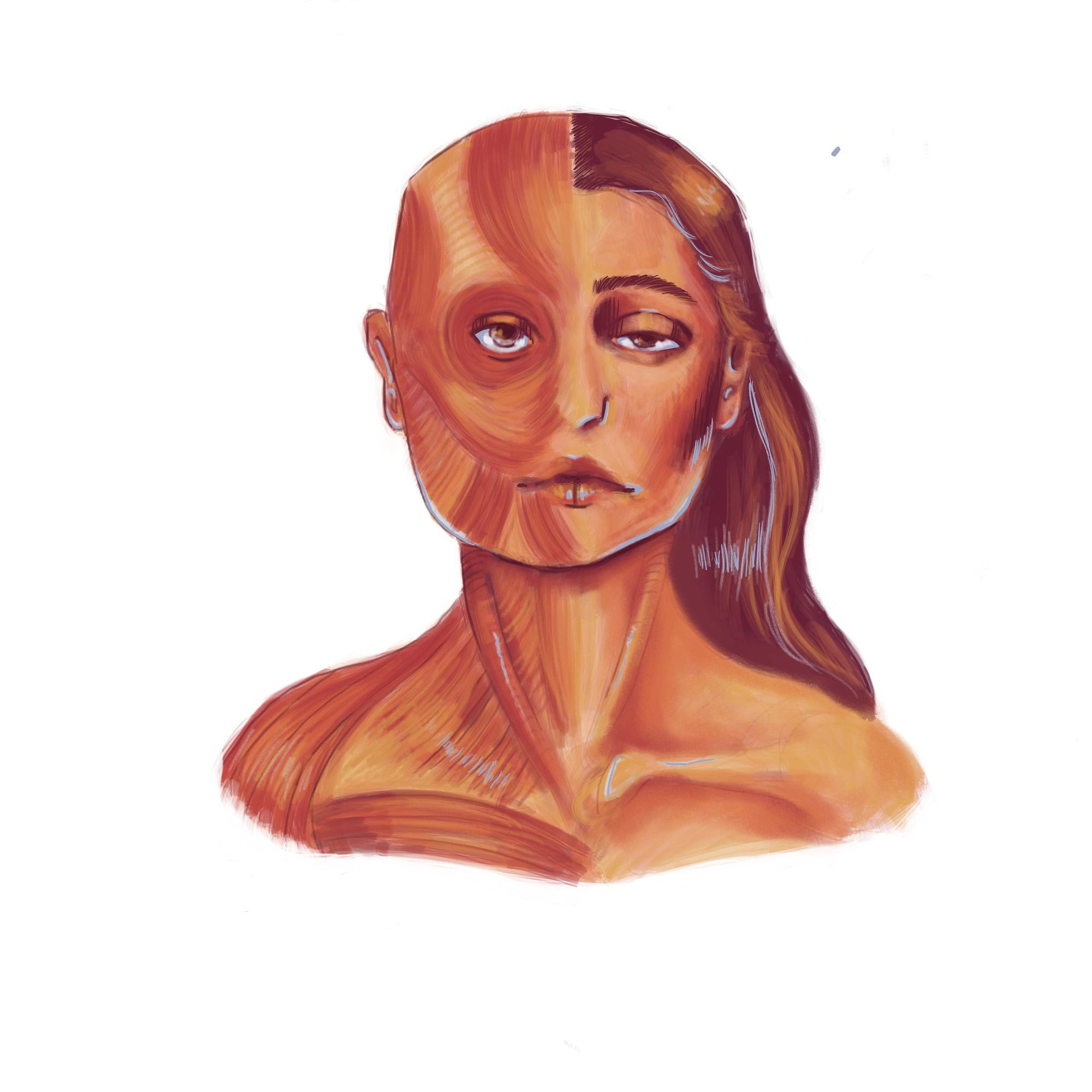
In the summer of 2021, new mom Hannah Ensor suddenly noticed facial weakness - she was unable to chew, drink out of a straw, or even smile [1]. “I had just eaten dinner, and we wanted to take some pictures on the pier during the sunset. I tried so hard to smile… And no matter what I did my facial muscles just wouldn’t cooperate,” Ensor said. Soon after, she began developing hand, wrist, and finger weakness. For a while, Ensor feared that she had multiple sclerosis. Tests and MRIs later ruled that out, but it was still unclear what the root cause of these symptoms was.
In March 2022, after multiple blood tests, Ensor received her diagnosis: myasthenia gravis (MG). The following month, she was prescribed medication and underwent surgery to remove her thymus, a small gland located between the lungs that is involved in the progression of MG. However, Ensor, who is impacted by fatigue and muscle weakness to this day, is just one of many victims of MG [1].
Ensor's story reflects the wider reality of myasthenia gravis. In this chronic autoimmune disorder, the body’s immune system mistakenly destroys muscle-nerve communication pathways in the neuromuscular junction, the connection site between a muscle and nerve [2]. This disruption of critical communication routes causes muscle weakness, particularly in the eyes and face, leading to difficulty in numerous facial functions [3]. Myasthenia gravis occurs in two forms: ocular MG and generalized MG [4]. In ocular MG, muscle weakness is restricted to the eyes, whereas in generalized MG, weakness arises in the eye, limb, and respiratory muscles [2]. About 51% of MG patients are initially diagnosed with ocular MG, and 55% of these cases convert to generalized MG later [5].
Although MG is a rare disease in and of itself, it is the most prevalent neuromuscular junction (NMJ) disorder, affecting about 37 per 100,000 people [6]. While MG research is limited by the lack of case studies, there are promising RNA treatments in clinical trials [7]. Additionally, epidemiological trends and risk factors associated with the disease are beginning to be identified [7].
II. History and Epidemiology
Myasthenia gravis was first identified by English physician Thomas Willis in 1672, but the disorder wasn’t distinctly classified as MG until much later [8]. In 1879, German physician Wilhelm Heinrich Erb differentiated MG from progressive bulbar palsy, a neuron deterioration disorder causing facial muscle weakness [9]. Later, in 1895, German neurologist Friedrich Jolly coined the term myasthenia gravis pseudoparalytica – severe muscle weakness with symptoms of paralysis [9].
The number of patients diagnosed with MG has consistently risen in the last few decades due to improved detection methods [10]. The annual average number of new MG cases per 100,000 has risen from 2.2 to 5.4 to 11.2 to 18 in each decade since 1980 [10]. Certain characteristics, such as age, ethnicity, and gender, may play a role in the likelihood of being diagnosed with MG [7].
The age distribution of those with MG has two primary trends: the disorder is more frequently seen in women aged 20-30 and in men aged over 50 [11]. Patients are generally classified into three subgroups based on their age at the onset of MG: early-onset MG when diagnosed under 50, late-onset MG when diagnosed from 50 to 64, and very-late-onset MG when diagnosed over 64 [12]. The highest percentage of patients diagnosed with MG are over 64, which may be due to increased life expectancy and improved disorder detection among older people. Patients with MG can face different symptoms based on when they were diagnosed with the disease [12].
Ethnicity is also observed to influence MG diagnosis. Anti-AChR antibodies, which play a major role in MG, are more frequent among Caucasians (71%) compared to African Americans (59%) [13]. However, African Americans face higher rates of early-onset MG and ocular MG compared to Caucasians [13]. In another study consisting of the Israeli Jewish population, ocular MG was more prevalent in those of Ashkenazi ethnicity, as opposed to those of non-Ashkenazi ethnicity [14]. In general, ethnic discrepancies in MG diagnosis and symptoms are likely due to underlying genetic differences among racial groups [13].
Sex is another risk factor for autoimmune diseases such as MG [15]. MG is more common in women than men, with incidence rates being 0.76 per 100,000 females and 0.60 per 100,000 males [16]. This may be due to a link between autoimmune diseases and the X chromosome, as females typically have two X chromosomes and males only have one X chromosome [15].
While ethnicity, age, and gender may impact MG diagnosis, more epidemiological data about the incidence and patterns of the disease is necessary to confirm these predictions [17].
III. Causes and Biological Basis
MG is typically caused by two factors: autoantibodies and thymus histology.
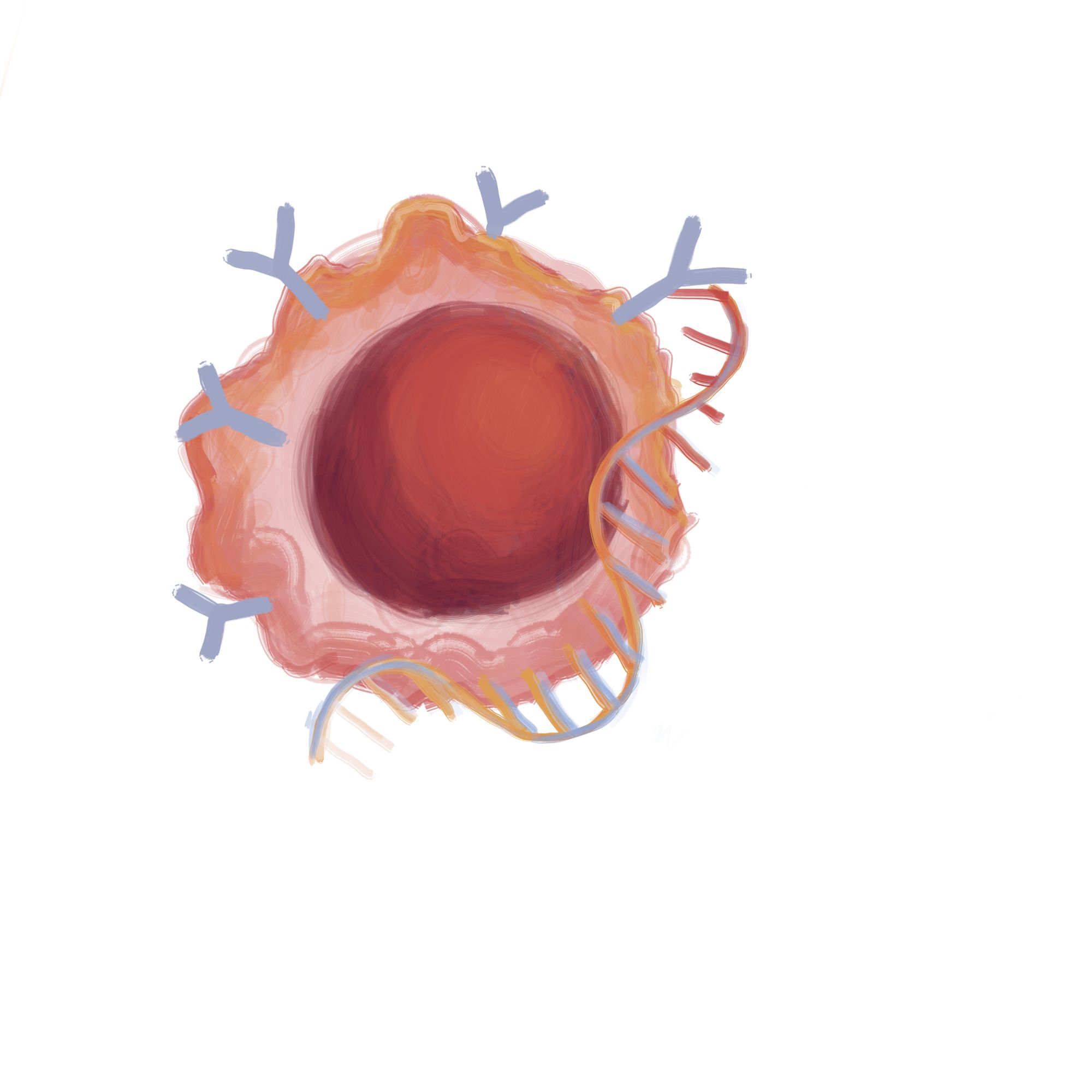
Normally, the brain sends electrical signals along neurons, which are transmitted to the muscle through neurotransmitters in the neuromuscular junction [18]. In healthy individuals, a neurotransmitter called acetylcholine binds to sites on skeletal muscles called acetylcholine receptors, activating muscle contraction [19]. In MG, however, anti-acetylcholine receptor (anti-AChR) antibodies block, alter, or destroy acetylcholine receptors, preventing muscle contraction [20][21]. While these self-harming antibodies, called autoantibodies, usually specifically block acetylcholine receptors, some also hinder other proteins involved in muscle contraction [22].

Abnormalities in the thymus gland, which produces T-cells that fight infections, are another cause of MG [23]. Normally, the thymus grows until puberty and then begins to shrink [24]. However, those with MG continue to have a large thymus after puberty, leading to thymic hyperplasia, which is an enlarged thymus, or thymomas, which are T-cell clusters that may result in tumors [24][25]. An enlarged thymus and cancerous tumors begin producing autoantibodies that attack the body’s cells and block acetylcholine receptors, ultimately disrupting neuromuscular transmission [24] [26]. Thymic hyperplasia is seen in approximately 38.2% of MG patients, while thymomas are found in about 36.4% of MG patients [24].
While the genetic origin of the disorder is still being investigated, researchers have identified multiple chromosomes that may be associated with MG [27]. However, inheriting MG is rare; only 3.8% - 7.1% of patients have a family history of the disease. Scientists have also found distinct genes impacting early and late-onset MG [27]. Additionally, in some cases, a pregnant woman may transfer autoantibodies to the fetus, but this disorder, known as neonatal MG, is generally temporary and lasts for about four months after birth [28].
IV. Symptoms, Progression, and Complications
Common symptoms of myasthenia gravis include eye muscle weakness, eyelid drooping, and blurred or double vision [30]. The patient may also encounter difficulty swallowing, impaired speech, facial drooping, and weakness in the limbs [30]. In some cases, weakness extends to breathing muscles and causes critical respiratory failure [30]. However, for the majority of patients, MG is debilitating but not fatal, with the disorder’s mortality rate being 1.4 per one million [31].
MG is a progressive disorder that spreads to the eye, limb, neck, and respiratory muscles approximately 4.5 months after onset [32]. Those with MG have a relatively favorable prognosis, but the lifespans of patients tend to be shorter than average [31].
The primary complication of MG is myasthenic crisis, a form of severe respiratory muscle weakness that leads to extreme breathing difficulty [33]. About 20% of those with MG face a myasthenic crisis within the first year of onset, and it is frequent in the first two years [33]. Frequent causes of a myasthenic crisis include underlying respiratory tract infection, inadequate MG treatment, and drug withdrawal [34]. A myasthenic crisis is addressed with immediate ventilator assistance or a non-invasive bilevel positive airway pressure (BiPAP) device, which is essentially a ventilation-assistance face mask [33][35].
V. Diagnosis
Early detection of myasthenia gravis is critical to managing the condition [37].
One method to test for MG is edrophonium testing, also known as a Tensilon test [37]. During this test, a medication called edrophonium is administered intravenously, through the vein, to a patient [37]. Edrophonium inhibits the acetylcholinesterase enzyme from breaking down acetylcholine in the NMJ, increasing acetylcholine concentration and allowing muscle contraction [38]. If a patient’s muscles become stronger after taking edrophonium, they initially did not have a high enough functional acetylcholine concentration and can be diagnosed with MG [38][39]. It should be noted that the FDA discontinued edrophonium testing in the United States in 2018 due to its high rate of false-positive results [40].
Another testing method is a repetitive nerve stimulation test [41]. This electrophysiological test measures the strength of electrical impulses sent along a nerve in response to a stimulus. If the impulse’s strength decreases by 10% or more by the fourth or fifth stimuli, muscle-nerve signaling is impaired and the patient may have MG [41]. However, the repetitive nerve stimulation test alone is not reliable for MG diagnosis and should be conducted alongside other diagnostic tests [42].
A third testing method to diagnose MG is the acetylcholine receptor (AChR) antibody test [21]. Anti-AChR antibodies prevent muscle contraction by disrupting acetylcholine from binding to AChR sites. Hence, the presence of three types of anti-AChR antibodies may be tested: those that block, modify, and most commonly, bind to AChR sites. High levels of these antibodies in blood serum can be used to diagnose MG [21].
VI. Treatment and Research
While there is no known cure for myasthenia gravis, approximately 35% of patients achieve long-term remission and live in a stable condition with no necessary use of medication [43]. For other patients, a common first-line medication prescribed is pyridostigmine, which is an acetylcholinesterase inhibitor that prevents acetylcholine breakdown [44]. A thymectomy, which is a thymus removal surgery, may be recommended to relieve MG symptoms [45].
Currently, RNA-based chimeric antigen receptor (CAR) T-cell therapy is in early-phase trials and demonstrates promising clinical results [46]. The CAR molecule can recognize, target, and bind to specific proteins, proving useful in treating autoimmune diseases like MG. Researchers from a biopharmaceutical company called Cartesian Therapeutics attempted to use RNA engineering to generate CAR T-cells that address antibody overproduction. In the study, RNA-based CAR T-cells called Descartes-08 were intravenously administered to 14 patients with MG. Outcomes were measured through four metrics related to quality of life and symptoms. The results show that the treatment was generally well-tolerated and clinically significant in reducing MG severity for four to nine months after administration [46].
Another RNA-based treatment currently in trial, called ALN-CC5, uses a small interfering RNA to silence the expression of a complement in the liver [47]. The complement system, which is a part of the immune system, enhances the function of antibodies, including the anti-AChR antibodies involved in MG [48]. A study by Kusner and colleagues found that a component of the mentioned liver complement, called C5, is involved in activating the complement system and inhibiting the NMJ [47]. The researchers tested a new treatment and found that in two wild-type rat species, silencing C5 via ALN-CC5 prevented NMJ inhibition and significantly improved MG symptoms for up to 71 days. Pre-clinical studies support the future development of ALN-CC5 as a potential therapeutic of MG in humans [47].
Apart from medication and surgery, MG patients can also improve their health through physical, psychological, respiratory, and balance training [49]. In one study, patients who underwent physical and psychological therapy showed significant decreases in fatigue [49]. Additionally, after a home-based respiratory muscle endurance program involving aerobic and resistance training, patients showed increased respiratory endurance and total ventilatory volume [49][50]. Balance strategy training also proves beneficial, with MG patients seeing over a 15% increase in functional ability, balance, and cognition [49].
VII. Conclusion
The journey of researching myasthenia gravis has revealed the intricacies and complexities of this disorder. Cases such as Hannah Ensor’s exemplify the challenges faced by those living with MG.
Although the disease is rare, incidences of MG have risen in recent decades due to improved diagnosis methods. Additionally, epidemiological trends related to the disorder have emerged, with age, ethnicity, and gender as risk factors. MG primarily impacts women aged 20-30 and men over 50, and also displays distinct ethnic, sex, and genetic trends.
Myasthenia gravis is primarily caused by anti-AChR antibodies and thymus irregularities. In those with the disorder, nerve signals to muscles are disrupted in the NMJ and eventually lead to muscle weakness. However, MG is generally debilitating and not fatal.
Diagnostic methods for myasthenia gravis include edrophonium testing, repetitive nerve stimulation, and anti-AChR antibody testing. Although there is currently no cure for MG, ongoing research supports promising RNA-based therapies in the future. Additionally, medication and surgery are options available to help reduce disease severity and improve lifestyle.
While a cure for myasthenia gravis remains elusive, the collective effort of medical professionals, researchers, and patients contributes to our evolving understanding of this disorder and brings us closer to a remedy.
VIII. References
[1] Ensor, H. (2023, October 10). My MG Story: Getting My Smile Back. Living with MG Stories; Myasthenia Gravis Foundation of America. Retrieved from https://myasthenia.org/MG-Community/Blog/my-mg-story-getting-my-smile-back
[2] De Roxas, R. C., Bagnas, M. A., Baldonado, J. J., Rivera, J. P., & Roxas, A. A. (2016). Clinical Profile and Outcome of Postthymectomy versus Non-Thymectomy Myasthenia Gravis Patients in the Philippine General Hospital: A 6-Year Retrospective Study. Frontiers in neurology, 7, 96. https://doi.org/10.3389/fneur.2016.00096
[3] Fatehi, F., Moradi, K., Okhovat, A. A., Shojatalab, G., Sedighi, B., Boostani, R., Sarraf, P., Haghi Ashtiani, B., Ghasemi, M., Moussavi, S., Anjidani, N., & Nafissi, S. (2021). Zytux in refractory myasthenia gravis: A multicenter, open-labeled, clinical trial study of effectiveness and safety of a rituximab biosimilar. Frontiers in Neurology, 12, 682622. https://doi.org/10.3389/fneur.2021.682622
[4] Yang, L., Tang, Y., He, F., Zhang, C., Kessi, M., Peng, J., & Yin, F. (2022). Clinical characteristics and outcome predictors of a Chinese childhood-onset myasthenia gravis cohort. Frontiers in Pediatrics, 10, 996213. https://doi.org/10.3389/fped.2022.996213
[5] Hendricks, T. M., Bhatti, M. T., Hodge, D. O., & Chen, J. J. (2019). Incidence, epidemiology, and transformation of ocular myasthenia gravis: A population-based study. American Journal of Ophthalmology, 205, 99–105. https://doi.org/10.1016/j.ajo.2019.04.017
[6] Rodrigues, E., Umeh, E., Aishwarya, Navaratnarajah, N., Cole, A., & Moy, K. (2024). Incidence and prevalence of myasthenia gravis in the United States: A claims-based analysis. Muscle & nerve, 69(2), 166–171. https://doi.org/10.1002/mus.28006
[7] Narayanaswami, P., Sanders, D. B., Wolfe, G., Benatar, M., Cea, G., Evoli, A., Gilhus, N. E., Illa, I., Kuntz, N. L., Massey, J., Melms, A., Murai, H., Nicolle, M., Palace, J., Richman, D., & Verschuuren, J. (2021). International consensus guidance for management of myasthenia gravis: 2020 update. Neurology, 96(3), 114–122. https://doi.org/10.1212/WNL.0000000000011124
[8] Hughes, T. (2005). The early history of myasthenia gravis. Neuromuscular Disorders: NMD, 15(12), 878–886. https://doi.org/10.1016/j.nmd.2005.08.007
[9] Keesey, J. (1998). Myasthenia gravis. Archives of Neurology, 55(5), 745–746. https://doi.org/10.1001/archneur.55.5.745
[10] Rutledge, S., Kenny, O., O'Riordan, S., McGuigan, C., & Tubridy, N. (2016). Myasthenia gravis: A population-based epidemiological study. Irish Medical Journal, 109(2), 355.
[11] Andersen, J. B., Heldal, A. T., Engeland, A., & Gilhus, N. E. (2014). Myasthenia gravis epidemiology in a national cohort; combining multiple disease registries. Acta Neurologica Scandinavica. Supplementum, (198), 26–31. https://doi.org/10.1111/ane.12233
[12] Cortés-Vicente, E., Álvarez-Velasco, R., Segovia, S., Paradas, C., Casasnovas, C., Guerrero-Sola, A., Pardo, J., Ramos-Fransi, A., Sevilla, T., López de Munain, A., Gómez, M. T., Jericó, I., Gutiérrez-Gutiérrez, G., Pelayo-Negro, A. L., Martín, M. A., Mendoza, M. D., Morís, G., Rojas-Garcia, R., Díaz-Manera, J., Querol, L., … Illa, I. (2020). Clinical and therapeutic features of myasthenia gravis in adults based on age at onset. Neurology, 94(11), e1171–e1180. https://doi.org/10.1212/WNL.0000000000008903
[13] Abukhalil, F., Mehta, B., Saito, E., Mehta, S., & McMurtray, A. (2015). Gender and ethnicity based differences in clinical and laboratory features of myasthenia gravis. Autoimmune Diseases, 2015, 197893. https://doi.org/10.1155/2015/197893
[14] Asmail, A., Kesler, A., Drory, V. E., Kolb, H., & Karni, A. (2017). Effect of ethnic origin and gender on the clinical manifestations of myasthenia gravis among the Jewish population in Israel. Journal of Neuroimmunology, 307, 47–52. https://doi.org/10.1016/j.jneuroim.2017.04.003
[15] Nicolì, V., Tabano, S. M., Colapietro, P., Maestri, M., Ricciardi, R., Stoccoro, A., Fontana, L., Guida, M., Miozzo, M., Coppedè, F., & Migliore, L. (2022). Preferential X chromosome inactivation as a mechanism to explain female preponderance in myasthenia gravis. Genes, 13(4), 696. https://doi.org/10.3390/genes13040696
[16] Chen, J., Tian, D. C., Zhang, C., Li, Z., Zhai, Y., Xiu, Y., Gu, H., Li, H., Wang, Y., & Shi, F. D. (2020). Incidence, mortality, and economic burden of myasthenia gravis in China: A nationwide population-based study. The Lancet Regional Health. Western Pacific, 5, 100063. https://doi.org/10.1016/j.lanwpc.2020.100063
[17] Carr, A. S., Cardwell, C. R., McCarron, P. O., & McConville, J. (2010). A systematic review of population based epidemiological studies in myasthenia gravis. BMC Neurology, 10, 46. https://doi.org/10.1186/1471-2377-10-46
[18] Deschenes, M. R., Patek, L. G., Trebelhorn, A. M., High, M. C., & Flannery, R. E. (2021). Juvenile neuromuscular systems show amplified disturbance to muscle unloading. Frontiers in Physiology, 12, 754052. https://doi.org/10.3389/fphys.2021.754052
[19] Zholos, A. V., Bolton, T. B., Dresvyannikov, A. V., et al. (2004). Cholinergic excitation of smooth muscles: Multiple signaling pathways linking M2 and M3 muscarinic receptors to cationic channels. Neurophysiology, 36, 398–406. https://doi.org/10.1007/s11062-005-0034-2
[20] Limburg, P. C., The, T. H., Hummel-Tappel, E., & Oosterhuis, H. J. (1983). Anti-acetylcholine receptor antibodies in myasthenia gravis. Part 1. Relation to clinical parameters in 250 patients. Journal of the Neurological Sciences, 58(3), 357–370. https://doi.org/10.1016/0022-510x(83)90095-3
[21] Peeler, C. E., De Lott, L. B., Nagia, L., Lemos, J., Eggenberger, E. R., & Cornblath, W. T. (2015). Clinical utility of acetylcholine receptor antibody testing in ocular myasthenia gravis. JAMA Neurology, 72(10), 1170–1174. https://doi.org/10.1001/jamaneurol.2015.1444
[22] Baggi, F., Andreetta, F., Maggi, L., Confalonieri, P., Morandi, L., Salerno, F., Bernasconi, P., Montomoli, C., Barberis, M., Mantegazza, R., & Antozzi, C. (2013). Complete stable remission and autoantibody specificity in myasthenia gravis. Neurology, 80(2), 188–195. https://doi.org/10.1212/WNL.0b013e31827b907b
[23] Melms, A., Schalke, B. C., Kirchner, T., Müller-Hermelink, H. K., Albert, E., & Wekerle, H. (1988). Thymus in myasthenia gravis. Isolation of T-lymphocyte lines specific for the nicotinic acetylcholine receptor from thymuses of myasthenic patients. The Journal of Clinical Investigation, 81(3), 902–908. https://doi.org/10.1172/JCI113401
[24] Tireli, H., Yuksel, G., Okay, T., & Tutkavul, K. (2020). Role of thymus on prognosis of myasthenia gravis in Turkish population. Northern Clinics of Istanbul, 7(5), 452–459. https://doi.org/10.14744/nci.2020.51333
[25] Zou, D., Luo, H., Feng, Y., Zeng, B., & Lei, Y. (2018). Massive thymic hyperplasia in an adult: A rare case report and literature review. International Journal of Surgery Case Reports, 47, 104–108. https://doi.org/10.1016/j.ijscr.2018.04.037
[26] Yamada, Y., Weis, C. A., Thelen, J., Sticht, C., Schalke, B., Ströbel, P., & Marx, A. (2020). Thymoma associated myasthenia gravis (TAMG): Differential expression of functional pathways in relation to MG status in different thymoma histotypes. Frontiers in Immunology, 11, 664. https://doi.org/10.3389/fimmu.2020.00664
[27] Renton, A. E., Pliner, H. A., Provenzano, C., Evoli, A., Ricciardi, R., Nalls, M. A., Marangi, G., Abramzon, Y., Arepalli, S., Chong, S., Hernandez, D. G., Johnson, J. O., Bartoccioni, E., Scuderi, F., Maestri, M., Gibbs, J. R., Errichiello, E., Chiò, A., Restagno, G., Sabatelli, M., … Traynor, B. J. (2015). A genome-wide association study of myasthenia gravis. JAMA Neurology, 72(4), 396–404. https://doi.org/10.1001/jamaneurol.2014.4103
[28] Kochhar, P. K., Schumacher, R. E., & Sarkar, S. (2021). Transient neonatal myasthenia gravis: Refining risk estimate for infants born to women with myasthenia gravis. Journal of Perinatology, 41(9), 2279–2283. https://doi.org/10.1038/s41372-021-00970-6
[29] Pal, S., & Sanyal, D. (2011). Jaw muscle weakness: A differential indicator of neuromuscular weakness--preliminary observations. Muscle & Nerve, 43(6), 807–811. https://doi.org/10.1002/mus.21990
[30] Cleanthous, S., Mork, A. C., Regnault, A., Cano, S., Kaminski, H. J., & Morel, T. (2021). Development of the Myasthenia Gravis (MG) Symptoms PRO: A case study of a patient-centred outcome measure in rare disease. Orphanet Journal of Rare Diseases, 16(1), 457. https://doi.org/10.1186/s13023-021-02064-0
[31] Christensen, P. B., Jensen, T. S., Tsiropoulos, I., Sørensen, T., Kjaer, M., Højer-Pedersen, E., Rasmussen, M. J., & Lehfeldt, E. (1998). Mortality and survival in myasthenia gravis: a Danish population based study. Journal of neurology, neurosurgery, and psychiatry, 64(1), 78–83. https://doi.org/10.1136/jnnp.64.1.78
[32] Zhou, Y., Chen, J., Li, Z., Tan, S., Yan, C., Luo, S., Zhou, L., Song, J., Huan, X., Wang, Y., Zhao, C., Zeng, W., & Xi, J. (2022). Clinical features of myasthenia gravis with antibodies to MuSK based on age at onset: A multicenter retrospective study in China. Frontiers in Neurology, 13, 879261. https://doi.org/10.3389/fneur.2022.879261
[33] Murthy, J. M., Meena, A. K., Chowdary, G. V., & Naryanan, J. T. (2005). Myasthenic crisis: Clinical features, complications and mortality. Neurology India, 53(1), 37–40. https://doi.org/10.4103/0028-3886.15050
[34] Sharma, S., Lal, V., Prabhakar, S., & Agarwal, R. (2013). Clinical profile and outcome of myasthenic crisis in a tertiary care hospital: A prospective study. Annals of Indian Academy of Neurology, 16(2), 203–207. https://doi.org/10.4103/0972-2327.112466
[35] Seneviratne, J., Mandrekar, J., Wijdicks, E. F., & Rabinstein, A. A. (2008). Noninvasive ventilation in myasthenic crisis. Archives of Neurology, 65(1), 54–58. https://doi.org/10.1001/archneurol.2007.1
[36] Sobierajski, T., Lasek-Bal, A., Krzystanek, M., & Gilhus, N. E. (2023). Diagnosis and therapy of myasthenia gravis-the patients' perspective: A cross-sectional study. Frontiers in Neurology, 14, 1214041. https://doi.org/10.3389/fneur.2023.1214041
[37] Domanovits, H., Wenger, S., Schillinger, M., Mayr, N., Holzer, M., Laggner, A. N., & Zeitlhofer, J. (2000). Der Edrophonium-Chlorid (Tensilon)-Test: Ein sichere Methode in der Diagnostik der Myasthenia gravis [Edrophonium chloride (Tensilon) test: a safe method in diagnosing myasthenia gravis]. Wiener Klinische Wochenschrift, 112(13), 592–595.
[38] Warnecke, T., Teismann, I., Zimmermann, J., Oelenberg, S., Ringelstein, E. B., & Dziewas, R. (2008). Fiberoptic endoscopic evaluation of swallowing with simultaneous Tensilon application in diagnosis and therapy of myasthenia gravis. Journal of Neurology, 255(2), 224–230. https://doi.org/10.1007/s00415-008-0664-6
[39] Katz, B., & Thesleff, S. (1957). The interaction between edrophonium (tensilon) and acetylcholine at the motor end-plate. British Journal of Pharmacology and Chemotherapy, 12(2), 260–264. https://doi.org/10.1111/j.1476-5381.1957.tb00131.x
[40] Naji, A., & Owens, M. L. (2023). Edrophonium. In StatPearls [Internet]. Treasure Island (FL): StatPearls Publishing. Retrieved from https://www.ncbi.nlm.nih.gov/books/NBK554566/
[41] Tomschik, M., Renaud, E., Jäger, F., Paternostro, C., Rath, J., Rinner, W., Zulehner, G., Zimprich, F., & Cetin, H. (2023). The diagnostic and prognostic utility of repetitive nerve stimulation in patients with myasthenia gravis. Scientific Reports, 13(1), 2985. https://doi.org/10.1038/s41598-023-30154-5
[42] Lo, Y. L., Najjar, R. P., Teo, K. Y., Tow, S. L., Loo, J. L., & Milea, D. (2017). A reappraisal of diagnostic tests for myasthenia gravis in a large Asian cohort. Journal of the Neurological Sciences, 376, 153–158. https://doi.org/10.1016/j.jns.2017.03.016
[43] Mano, K., Takegami, T., Okamoto, S., & Takahashi, A. (1993). Remission of myasthenia gravis: Clinical, electrophysiological and immunological studies. Nagoya Journal of Medical Science, 55(1-4), 103–113.
[44] Remijn-Nelissen, L., Verschuuren, J. J. G. M., & Tannemaat, M. R. (2022). The effectiveness and side effects of pyridostigmine in the treatment of myasthenia gravis: A cross-sectional study. Neuromuscular Disorders: NMD, 32(10), 790–799. https://doi.org/10.1016/j.nmd.2022.09.002
[45] Wolfe, G. I., Kaminski, H. J., Aban, I. B., Minisman, G., Kuo, H. C., Marx, A., Ströbel, P., Mazia, C., Oger, J., Cea, J. G., Heckmann, J. M., Evoli, A., Nix, W., Ciafaloni, E., Antonini, G., Witoonpanich, R., King, J. O., Beydoun, S. R., Chalk, C. H., Barboi, A. C., … MGTX Study Group. (2016). Randomized trial of thymectomy in myasthenia gravis. The New England Journal of Medicine, 375(6), 511–522. https://doi.org/10.1056/NEJMoa1602489
[46] Granit, V., Benatar, M., Kurtoglu, M., Miljković, M. D., Chahin, N., Sahagian, G., Feinberg, M. H., Slansky, A., Vu, T., Jewell, C. M., Singer, M. S., Kalayoglu, M. V., Howard, J. F., Jr, Mozaffar, T., & MG-001 Study Team (2023). Safety and clinical activity of autologous RNA chimeric antigen receptor T-cell therapy in myasthenia gravis (MG-001): a prospective, multicentre, open-label, non-randomised phase 1b/2a study. The Lancet. Neurology, 22(7), 578–590. https://doi.org/10.1016/S1474-4422(23)00194-1
[47] Kusner, L. L., Yucius, K., Sengupta, M., Sprague, A. G., Desai, D., Nguyen, T., Charisse, K., Kuchimanchi, S., Kallanthottathil, R., Fitzgerald, K., Kaminski, H. J., & Borodovsky, A. (2019). Investigational RNAi therapeutic targeting C5 is efficacious in pre-clinical models of myasthenia gravis. Molecular Therapy. Methods & Clinical Development, 13, 484–492. https://doi.org/10.1016/j.omtm.2019.04.009
[48] Iacomino, N., Vanoli, F., Frangiamore, R., Ballardini, M., Scandiffio, L., Bortone, F., Andreetta, F., Baggi, F., Bernasconi, P., Antozzi, C., Cavalcante, P., & Mantegazza, R. (2022). Complement activation profile in myasthenia gravis patients: Perspectives for tailoring anti-complement therapy. Biomedicines, 10(6), 1360. https://doi.org/10.3390/biomedicines10061360
[49] Corrado, B., Giardulli, B., & Costa, M. (2020). Evidence-based practice in rehabilitation of myasthenia gravis. A systematic review of the literature. Journal of Functional Morphology and Kinesiology, 5(4), 71. https://doi.org/10.3390/jfmk5040071
[50] Westerberg, E., Molin, C. J., Lindblad, I., Emtner, M., & Punga, A. R. (2017). Physical exercise in myasthenia gravis is safe and improves neuromuscular parameters and physical performance-based measures: A pilot study. Muscle & Nerve, 56(2), 207–214. https://doi.org/10.1002/mus.25493