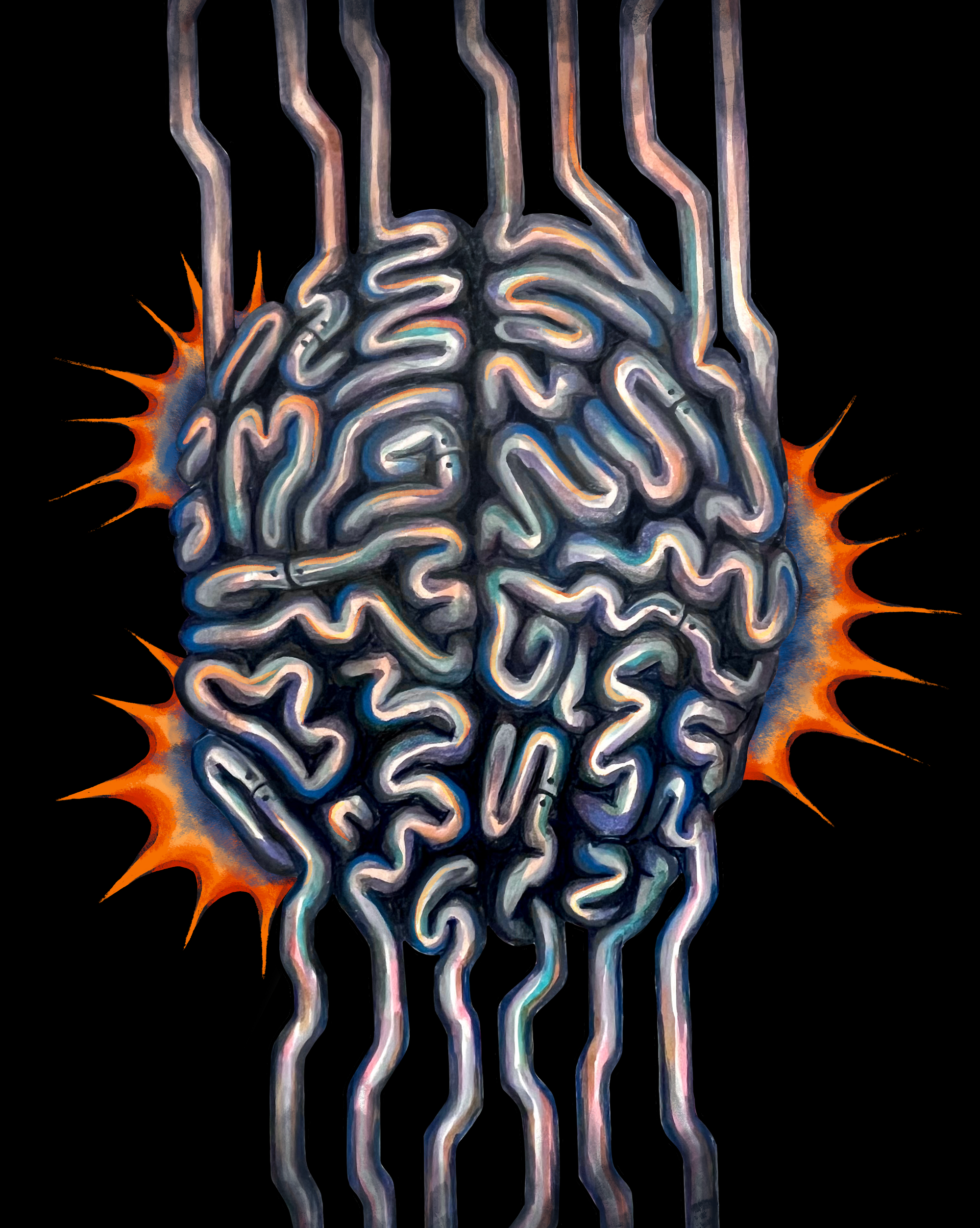
We are all familiar with the futuristic scenario where humans become one with machines to push beyond our biological limitations. Whether this idea excites or frightens you, it is currently becoming our reality. Developments in our understanding of the nervous system, along with constant refinements in neurotechnology, allow us to more directly and accurately interact with the central nervous system (CNS). By understanding our brain signals and electrically imitating those signals through different pieces of technology, we can trigger parts of the body the CNS can no longer reach to help restore functionality for impaired individuals [1].
Spinal cord injuries (SCIs) have profound, often life-altering consequences. The loss of crucial CNS communication pathways, which are responsible for movement, can result in paralysis [2]. Beyond functional loss, these injuries often result in serious psychological and emotional challenges. Transitioning from an independent life to one with physical limitations and an extensive need for outside help can be mentally taxing and lead to feelings of depression and hopelessness. These feelings can be exacerbated by societal stigma, inaccessibility, and misconceptions surrounding the injury, making it difficult for patients to reintegrate into society. In addition, treatment and management of SCIs carry substantial monetary costs, which can further burden both the patient and their family [2].
While one might assume that the tragic outcomes of SCIs come exclusively from the initial destruction of neural connections and blood vessels within the spinal cord, much of the damage occurs well after the initial injury [3]. The spinal cord’s response to the initial injury phase results in a secondary phase which continues for hours to days post-injury. Many factors stemming from the first stage such as vascular damage and unregulated release of neurotransmitters result in an unfavorable environment for cells within the nervous system including neurons, leading to their death. When this secondary phase persists, these factors become chronic and result in further neuronal death and the formation of glial scars. These scars block neurons from signaling to each other, making it very difficult to achieve functional recovery once the scars have fully formed [3].
For many decades, conventional clinical treatments for SCIs primarily focused on reducing the extent of initial injury. By stabilizing the spine and relieving pressure on the injured cord, doctors would minimize continued harm post-injury. This in turn would maximize the potential for functional recovery through rehabilitation and the body’s own ability to heal as it would from a scratch or broken bone [4]. While this offered positive results, standard treatments typically did not include external intervention to help reconnect these severed neural pathways. As a result, many patients had to endure the permanent functional loss sustained from their injury [4].
Despite innovations in the field of neuroscience regarding SCI treatment, translating these new findings into a widespread clinical setting remains challenging. This can be attributed to many obstacles, including a relatively small human subject pool, large differences between injury types, and substantial regulatory hurdles. But just as all science builds on itself, these previous advancements have built on one another to help develop a groundbreaking therapy: a Brain-Spine Interface (BSI). This digital bridge between the brain and spinal cord is changing the narrative of people affected by SCIs. By offering a chance to regain the movements we often take for granted, patients are given the hope to one day live independently [1].
Making a Move
This revolutionary achievement is the result of decades of dedicated research. To fully appreciate this breakthrough, we must first examine the scientific developments that ultimately made it possible, many of which lie no further than your middle school frog dissection.
While this may seem like an oversimplification, these experiments allowed us to understand the role of neurons in muscle contractions. Building on these principles, researchers began to use electrodes on the skin to conduct electricity, eliciting muscle movement in paralyzed patients through a technique known as Functional Electrical Stimulation (FES) [5]. Just like the frog experiment, external electrical stimuli from the researchers applied to the peripheral nerves worked to generate muscle contractions.
However, there was one large problem: the stimulation wasn’t controlled by the patient’s thoughts and was instead externally controlled. Due to the technological limitations, the models available only allowed patients to hold a remote and press buttons to elicit specific movements. In other words, none of their actions came directly from the brain. This is where another amazing development comes into play: Brain-Computer Interfaces (BCIs).
Brain and Machines
The name “Brain-Computer Interface” sounds futuristic, implying a device capable of linking mind and machine. However, BCIs have been tested on humans since the 1990s, albeit with large limitations on their capabilities. The purpose of this technology was to allow direct communication between the brain and external devices, which would be used to exchange or manipulate brain signals outside the body. Early BCIs were developed for individuals with severe motor disabilities, allowing them to communicate and control devices. From these initial BCIs, we have since developed less invasive and more accurate models.
With many signals coming from the brain, the process of filtering out relevant motor signals, which could be translated by a BCI into a controlled movement, proved extremely challenging. Years of research have overcome many of these challenges, resulting in the ability to reliably capture and decode information related to motor movement intentions. Researchers have used a wide variety of methods to read these “brain waves.” One particular study utilized magnetoencephalography (MEG) to capture these motor signals, measuring the magnetic fields generated by the electrical impulses of the brain. For this experiment, participants were told to physically move their wrist for one trial, and imagine doing it for another [6]. By cross-referencing the MEG recordings, the researchers concluded that there was a significant similarity between both overt and intended movement signals. This provided evidence that researchers can use MEG to accurately detect movement intentions, which can then be used as inputs for a BCI and for localizing the exact parts of the brain responsible for movement among different individuals [6].
Connecting BCIs to SCIs
When considering the best way to repair a rift in the spinal cord, which serves as the nervous system's highway, the obvious solution would be to directly fill the gap with more neurons. In reality, this has proven difficult because the area we hope to fill becomes overgrown with scar tissue, and new neurons often struggle to establish connections with the spinal cord [3]. As an alternative strategy, scientists are aiming to develop another approach to simply bypass the injury and build a bridge over the damaged section [7]. This would involve designing a BCI that could detect signals from the brain and then transmit these signals to either electrode arrays implanted in the spinal cord, eliciting movement of an actual arm, or to a prosthetic arm. Either of these types of BCIs would restore movement signals in the places where the CNS connection has been disrupted, showing great promise for bypassing injury sites [7].
The complexity of human physiology made it more feasible to start developing these therapies by connecting BCIs to a prosthetic arm rather than to transmit signals to restore actual arm movements, due to the prosthetics having fewer moving parts. In one study, scientists implanted microelectrodes in a participant’s brain and trained the BCI algorithm for several months by walking participants through various maneuvers [8]. Concurrently, the subject fine-tuned their control of the BCI prosthetic, just as one might fine-tune a new skill. By the end, the patient showed a large improvement in target-based reaching tasks and coordinated movements with the prosthetic limb [8].
While prosthetics showed great promise in improving the quality of life for people with disabilities such as those stemming from SCIs, the ultimate goal was always for the patient to regain control of their own body using their mind. The principle of using brain readings to control a prosthetic showed that in theory, one could use those same readings to trigger electrodes and stimulate specific paralyzed body parts through FES. However, connecting these two ideas to help treat paralysis required more testing to see if the concept held up in practice.

Bringing it Together
Using electrical stimulation in conjunction with BCIs to make a more natural recovery for paralyzed individuals requires us to start with basic model organisms. Experiments in rats resulted in the development of electrode placement protocols on the spine to mimic the natural activation of muscles [9]. These studies also gave rise to spinal implants that contained electrodes and control software that could target and modulate different muscle groups with better precision. As these stimulator technologies began to develop, arrays of electrodes could be directly implanted onto the spinal cord to increase the precision of stimulation [7]. As a result, there have been great developments in the degree of freedom patients are able to experience in their control of muscle movement.
This set the groundwork for researchers to begin treating SCIs in more compatible primate models using the same methods. In one particular study, the researchers extended this concept to non-human primates, or monkeys [10]. They implanted microelectrodes in the left motor cortex of the monkey’s brain, in an area responsible for leg movements. These microelectrodes were used to detect neural activity, which was then decoded with a computer. Using Bluetooth, these commands, or “motor states,” were then sent to an electric stimulator on the lower region of the spinal cord, which then allowed for wireless control of leg muscle movements. Even after partial tears of the spinal cord, this Brain-Spine Interface largely restored walking capabilities in the monkeys [10]. The methods used in this study have been approved for potential use in human research, paving the way for promising studies in humans with spinal cord injuries.
Bridging the Injury
The culmination of all this research is integration of these technologies into an implanted human-compatible brain-spine interface (BSI), one type of BCI. Doing so would help restore the participants’ control of their own body movements. This was first successful in a human in 2023. The study was conducted on a single patient, who was not only able to regain natural control over his movement, but also retain some of his neurological capabilities, even when the device was switched off [1]. This BSI builds off of existing electrical stimulation techniques, such as FES on the spine, by allowing fine-tuned control and the ability to modulate muscle activity in sync with motor intentions. One big innovation is that these devices can now communicate in quasi-real time.
To first understand which regions of the brain are responsible for motor movements in each individual patient, a surgeon uses computerized tomography and MEG to find the parts of the brain that respond most to movement intentions by having the patient imagine moving different parts of their body. The surgeon then operates to place the cortical implants in the corresponding locations on top of the brain. They also implant electrodes into the spine at locations that they have mapped to mobilization of muscle groups. They then calibrate these electrodes to determine an optimal amount of stimulation. By decoding the motor neural impulses coming from the brain, the electric cortical implants are able to send signals to bypass the injury via the Brain-Spine Interface and cause the desired movement in the legs.
Despite not experiencing the same amount of control with the device turned off, the patient was still able to walk independently with crutches, marking a substantial gain from his previous disability [1]. These extra gains are very promising, showing that this brain-controlled stimulation improves long-term locomotion. While the mechanisms behind this recovery aren’t well understood, studies with rats indicate that activating the local motor neurons may induce neuroplasticity, which can help reorganize the nerve cells to form new connections, allowing participants to bridge the injury site even when the BCI is turned off [11] [12].
This technology marks an exciting breakthrough in the treatment of motor deficits from neurological disorders such as SCI. Previous implants only allowed the patient to take several steps, but now he was able to walk impressive distances with the assistance of the BSI [1]. The BSI allowed for nearly twice as many steps within a given time period when compared to epidural electric stimulation, and also allowed for more natural walking patterns. It is important to note that the patient had partial spinal cord damage and the extent of recovery in each individual likely depends on the severity of the injury. With this being said, this has shown great promise for broad treatment of paralysis, as the patient was able to restore some motor function that he hadn’t had since before the injury [1].
Next Steps
As the technology behind the Brain-Spine Interface continues to improve through ongoing research, researchers are working to make the implants less invasive and more efficient, increasing accessibility of treatment and ultimately restoring the communication capabilities between the brain and the spine.
This technology marks a huge win in the treatment of SCIs, but it is not without shortcomings. Due to the signal processing between the brain, the device, and the spinal cord, there is latency between when the patient thinks of a movement and when the movement occurs [11]. Repairing the injury, rather than bypassing it, would ensure real-time communication between the brain and the spine, and is an ongoing and promising area of research.
In this pursuit of spinal cord repair, alternative strategies are being investigated, which also show great potential. Among these, stem cell therapy and the use of biomaterial scaffolds stand out as potentially transformative approaches. Stem cell treatments offer the possibility of replacing damaged neural tissue but still face many problems with regard to reintegrating the neurons into the existing neural network and removing glial scars [13]. Likewise, biomaterial scaffolds offer a way to regrow neurons by providing a supportive framework similar to the natural environment of the spinal cord [14]. By combining stem cell therapy with the use of biomaterial scaffolds, researchers aim to create an optimal microenvironment for neural regeneration.
While these and other approaches show promising results, they are still under active research and development, and they have not been used much in a clinical setting. This makes it even more important for us to appreciate the amazing progress that has been made through the Brain-Spine Interface and the hope it gives to patients with SCIs for regaining motor movement and improving their lives.
References
- Lorach, H., Galvez, A., Spagnolo, V., Martel, F., Karakas, S., Intering, N., Vat, M., Faivre, O., Harte, C., Komi, S., Ravier, J., Collin, T., Coquoz, L., Sakr, I., Baaklini, E., Hernandez-Charpak, S. D., Dumont, G., Buschman, R., Buse, N., Denison, T., … Courtine, G. (2023). Walking naturally after spinal cord injury using a brain-spine interface. Nature, 618(7963), 126–133. https://doi.org/10.1038/s41586-023-06094-5
- Budd, M. A., Gater, D. R., Jr, & Channell, I. (2022). Psychosocial Consequences of Spinal Cord Injury: A Narrative Review. Journal of personalized medicine, 12(7), 1178. https://doi.org/10.3390/jpm12071178
- Anjum, A., Yazid, M. D., Fauzi Daud, M., Idris, J., Ng, A. M. H., Selvi Naicker, A., Ismail, O. H. R., Athi Kumar, R. K., & Lokanathan, Y. (2020). Spinal Cord Injury: Pathophysiology, Multimolecular Interactions, and Underlying Recovery Mechanisms. International journal of molecular sciences, 21(20), 7533. https://doi.org/10.3390/ijms21207533
- Kwon, W.-K., Park, D.-H., Kim, H., Park, W.-B., Oh, J.-K., Moon, H. J., Kim, J. H., & Park, Y.-K. (2018). Surgical Treatment in Neurocritical Care for Acute Spinal Cord Injuries. Journal of Neurointensive Care, 1(1), 15-19. https://doi.org/10.32587/jnic.2018.00052
- Ho, C.H., Triolo, R.J., Elias, A.L., Kilgore, K.L., DiMarco, A.F., Bogie DPhil, K., Vette, A.H., Audu, M.L., Kobetic, R., Chang, S.R., Chan, K.M., Dukelow, S., Bourbeau, D.J., Brose, S.W., Gustafson, K.J., Kiss, Z.H.T. & Mushahwar, V.K. (2014). Functional electrical stimulation and spinal cord injury. Physical Medicine and Rehabilitation Clinics, 25(3), 631-654. https://doi.org/10.1016/j.pmr.2014.05.001
- Wang, W., Sudre, G. P., Xu, Y., Kass, R. E., Collinger, J. L., Degenhart, A. D., Bagic, A. I., & Weber, D. J. (2010). Decoding and cortical source localization for intended movement direction with MEG. Journal of neurophysiology, 104(5), 2451–2461. https://doi.org/10.1152/jn.00239.2010
- Calvert, J. S., Grahn, P. J., Strommen, J. A., Lavrov, I. A., Beck, L. A., Gill, M. L., Linde, M. B., Brown, D. A., Van Straaten, M. G., Veith, D. D., Lopez, C., Sayenko, D. G., Gerasimenko, Y. P., Edgerton, V. R., Zhao, K. D., & Lee, K. H. (2019). Electrophysiological Guidance of Epidural Electrode Array Implantation over the Human Lumbosacral Spinal Cord to Enable Motor Function after Chronic Paralysis. Journal of Neurotrauma, 36(9), 1451–1460. https://doi.org/10.1089/neu.2018.5921
- Collinger, J. L., Wodlinger, B., Downey, J. E., Wang, W., Tyler-Kabara, E. C., Weber, D. J., McMorland, A. J., Velliste, M., Boninger, M. L., & Schwartz, A. B. (2013). High-performance neuroprosthetic control by an individual with tetraplegia. Lancet (London, England), 381(9866), 557–564. https://doi.org/10.1016/S0140-6736(12)61816-9
- Wenger, N., Moraud, E. M., Gandar, J., Musienko, P., Capogrosso, M., Baud, L., Le Goff, C. G., Barraud, Q., Pavlova, N., Dominici, N., Minev, I. R., Asboth, L., Hirsch, A., Duis, S., Kreider, J., Mortera, A., Haverbeck, O., Kraus, S., Schmitz, F., DiGiovanna, J., … Courtine, G. (2016). Spatiotemporal neuromodulation therapies engaging muscle synergies improve motor control after spinal cord injury. Nature medicine, 22(2), 138–145. https://doi.org/10.1038/nm.4025
- Capogrosso, M., Milekovic, T., Borton, D., Wagner, F., Moraud, E. M., Mignardot, J. B., Buse, N., Gandar, J., Barraud, Q., Xing, D., Rey, E., Duis, S., Jianzhong, Y., Ko, W. K., Li, Q., Detemple, P., Denison, T., Micera, S., Bezard, E., Bloch, J., … Courtine, G. (2016). A brain-spine interface alleviating gait deficits after spinal cord injury in primates. Nature, 539(7628), 284–288. https://doi.org/10.1038/nature20118
- Bonizzato, M., Pidpruzhnykova, G., DiGiovanna, J., Shkorbatova, P., Pavlova, N., Micera, S., & Courtine, G. (2018). Brain-controlled modulation of spinal circuits improves recovery from spinal cord injury. Nature communications, 9(1), 3015. https://doi.org/10.1038/s41467-018-05282-6
- Tang, S., Cuellar, C. A., Song, P., Islam, R., Huang, C., Wen, H., Knudsen, B. E., Gong, P., Lok, U. W., Chen, S., & Lavrov, I. A. (2020). Changes in spinal cord hemodynamics reflect modulation of spinal network with different parameters of epidural stimulation. NeuroImage, 221, 117183. https://doi.org/10.1016/j.neuroimage.2020.117183
- Nakamura, M., & Okano, H. (2013). Cell transplantation therapies for spinal cord injury focusing on induced pluripotent stem cells. Cell research, 23(1), 70–80. https://doi.org/10.1038/cr.2012.171
- Guijarro-Belmar, A., Varone, A., Baltzer, M. R., Kataria, S., Tanriver-Ayder, E., Watzlawick, R., Sena, E., Cunningham, C. J., Rajnicek, A. M., Macleod, M., Huang, W., Currie, G. L., & McCann, S. K. (2022). Effectiveness of biomaterial-based combination strategies for spinal cord repair - a systematic review and meta-analysis of preclinical literature. Spinal cord, 60(12), 1041–1049. https://doi.org/10.1038/s41393-022-00811-z
