
Rare diseases and genetic mutations
News articles create beautified names for genetic disorders like the Butterfly Children (epidermolysis bullosa), Moon Children (albinism), Porcelain Doll (osteogenesis imperfecta), and Panda Baby (Gaucher disease), all attempting to hide the grotesque nature of the disorders behind glamorized euphemisms for better public acceptance. Yet the mysterious nature of the mechanisms behind many of those disorders remained unsolved until the first DNA analysis was conducted in the 1980s [1]. Among these mysteries was the complex phenomenon that puzzled scientists for many years: the “Puppet Children.”
In 1965, Dr. Harry Angelman documented cases of three children exhibiting sudden and uncontrollable laughter, intellectual impairment, abnormal facial features, jerky movements, and a protruding tongue, which was then collectively defined as Angelman syndrome (AS) [2]. As many as 1 in 12,000 people in the United States are afflicted by AS. Scientists soon identified that this rare neurogenetic disorder was caused by a maternal mutation (genetic information from their biological mother) of chromosome 15, which contains a crucial gene called UBE3A [2]. A gene is a segment of DNA that carries hereditary information needed to specify our biological traits. Deleterious mutations in the DNA sequence cause alterations in normal gene expression, leading to a reduction of the organism’s fitness in its current environment. In the neurons of normal individuals, the maternal allele of UBE3A is usually expressed and the paternal allele (genetic information from their biological father) is silenced. However, for AS patients, the loss of both UBE3A copies disrupts its function in synthesizing enzymes and forming neural connections in various brain areas [2].
The threats posed by deleterious mutations such as the UBE3A mutation on human health have pressured scientists to uncover the intricate mechanisms behind genetic disorders and gene editing. Given the complexity of human embryonic development, frontline researchers struggled to completely decipher the human genome and propose a cure to reverse these congenital deficits.
Clinical Overview: Case studies comparison
Other than leading to enzyme dysfunction, the loss of UBE3A eventually results in four paths of genetic mechanisms that lead to AS: deletion of the part of the UBE3A gene is from in 70–75% of patients, other mutations in the UBE3A gene in 10-15% of the patients, uniparental disomy in 2–3% of patients, and genomic imprinting defects in 3–5% of the patients. Each contributes to varying degrees of phenotype severity of AS [2].
Individuals experiencing AS can exhibit different symptoms in differing clinical severity, mostly determined by the four underlying mechanisms previously mentioned: deletion, mutation, uniparental disomy, and imprinting defects [3]. Uniparental disomy occurs when an error during mitosis or meiosis leads to individuals receiving both chromosomes of the same gene from one parent, and none from the other parent. In this case, AS patients inherited two copies of the silent UBE3A chromosome from the father. The lack of UBE3A expression can also result from genomic imprinting, a process regulated by the transcription of UBE3A-ATS, a DNA strand that silences the parental allele. Mistakes that occur during imprinting cause the maternal chromosome to carry the UBE3A-ATS sequence, preventing expression on both alleles. Patient 1 from the cases above demonstrates chromosome 15 section q11-13 deletion while Patient 2 exhibits imprinting defects. Overall, scientists identified that deletion patients are most likely to exhibit the complete set of AS symptoms–severe mental retardation, seizures, hyperactive behaviors, obesity, sleep disturbances, and more [3]. Mutation patients tend to have an intermediate phenotype and uniparental disomy patients usually experience even milder conditions. As a result of intellectual impairment and developmental delay, children with AS often fail to acquire adequate cognitive and motor skills [3].
Below are two case studies comparing a chromosome deletion patient to an imprinting defects patient.
Patient 1 is a five-year-old girl with hypotonia, a type of abnormal development that affects one’s ability to control muscles [4]. She could sit without support at 8 months and walk independently at 24 months, much later than the average 6 months and 12 months. Furthermore, she exhibited severe intellectual disability and restricted oral language skill limited to approximately 10 words. Some physical features of this patient include a protruding jaw, large tongue, wide mouth, and wide-spaced teeth. Her overall demeanor is happy and hyperactive, with frequent disrupted sleep cycles. By the age of 20 months, she has developed frequent drooling and early-onset seizures [4].
Patient 2 is a 17-year-old female with normal growth development [5]. Compared to Patient 1, she is able to sit at 6 months, walk at 12 months, run upstairs, and ride a bicycle without assistance. Her vocabulary includes over 100 words. Similar to Patient 1, she behaves happily with intermittent aggression when encountering difficulties in communicating. However, unlike Patient 1, Patient 2 experiences no seizures [5]. Disregarding the age gap, scientists conclude that AS Patient 2 displays milder symptoms than AS Patient 1, but why? Has Patient 2 received better treatment and reversed the damage of UBE3A loss?
AS in mice: neurological and molecular mechanisms
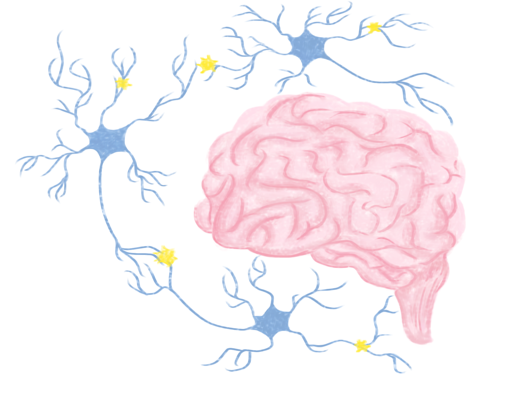
Mice have long been used as models for research for neurological diseases due to the similarity between their nervous system to that of humans, with the UBE3A gene sharing a similar location. Brains utilize cells called neurons to transport electrical signals called action potentials down an axon from the cell body of one neuron to the dendrites of another, building a network of “communication” in the brain. In between the axon and the dendrite are the synapse, a gap in which electrical or chemical (neurotransmitter) impulses pass from one neuron to the next. However, the initiation and inhibition of these action potentials are determined by a secondary signal that is stored in the dendritic spine, which receives and regulates the signals it receives from another neuron [6].
Investigating changes in AS mouse behavior has led to two clear effects: a significant reduction in dendritic spine density and length in adult mice and also changes in synaptic plasticity, or the capacity for neurons to strengthen their connections [7]. Reduction in dendritic spine density disrupts the transmission of electrical signals to other neurons. In AS mice, the loss of UBE3A results in the creation of silent synapses, where the initial signal fails to activate a proper postsynaptic response, consequently disrupting the normal circuitry flow [8]. Abnormal connectivity is likely caused by disabling synaptic plasticity and contributes to the measured decrease in dendritic spine density [9]. When neurons cannot effectively communicate with each other, UBE3A-deficient mouse models experience cognitive deficits in areas such as learning and memory [7]. This may also account for the cognitive deficits, speech, and motor impairments seen in AS patients.
Differential diagnosis
There are no signs of Angelman syndrome at birth, with symptoms usually emerging at around 6 to 12 months. Early-onset epilepsy appears in most AS patients from the age of 1-3 [8]. Patients suffer from either sudden, rapid, muscle contract movements, or slow, prolonged seizures, leading to a loss of consciousness. Non-deletion patients may have late-onset epilepsy, or be completely seizure-free [10]. The symptomatic variability makes it extremely difficult to diagnose AS patients during early infancy, especially when AS overlaps with other neurogenetic disorders that are also caused by small deletions or single-gene mutations [11].
Many clinically indistinguishable but genetically distinct cases are reflective of a convergent biology and common regions disrupted by mutations. The protein products of many genes that cause overlapping phenotypes interact directly or indirectly and function in coinciding pathways [12]. For example, both Angelman syndrome and Rett syndrome, caused by an X-linked single-gene mutation, exhibit symptoms like absent speech, microcephaly, stereotypic hand movements, happy dispositions, and seizures in early childhood [13]. The protein mutated in Rett syndrome plays a crucial role in stabilizing and maintaining mature neurons, and its absence reduces dendritic spine density and neuronal plasticity [13]. On the other hand, the enzyme synthesized by the UBE3A gene in AS has been identified to regulate protein expression by targeting other proteins to be degraded within cells [2]. The similar functions of these proteins suggest overlapping gene regulation pathways. Conditions like Mowat-Wilson syndrome and Pitt-Hopkins syndrome, which closely resemble AS in older children, are often associated with AS phenotypes both clinically and in terms of treatment. To add strength to these diagnoses of AS, the clinician could utilize additional technologies such as individual sequencing of a specific gene or whole-exome sequencing [11].
Genomic imprinting and future directions
Researchers have spent several years trying to find a cure for AS, but the nervous system pathologies of AS still remain elusive. Most current treatments focus on behavioral and drug therapies to alleviate each symptom: changing patients’ lifestyles to improve gastrointestinal issues, using medications to reduce seizures and anxiety, and sleep training. However, other drugs in development for AS aim to externally supply a new copy of the UBE3A gene into the brain. Through hippocampal injection of a virus that carries a copy of the UBE3A gene, the UBE3A-deficient mouse models are often able to restore the gene expression to normal levels [14]. The hippocampus is a part of the brain that is involved in is involved in memory, learning, and emotion. In one study, mice that underwent this gene replacement experiment significantly improved learning and memory compared to the control for up to 17 months after the first injection [15]. However, this treatment failed to reduce motor deficits in AS mice, with scientists suspecting that it was because the virus failed to reach the cerebellum, a part of the brain that coordinates movement and balance [15].
For gene therapy to be a viable treatment option, researchers have proposed strategies for direct restoration of the UBE3A gene. Antisense oligonucleotides (ASOs) are single-stranded nucleic acid molecules structurally similar to RNA. When binding to RNA, they can approach the nucleus to break down the UBE3A-ATS, un-silencing the parental UBE3A gene in adult mice [16]. Though the mice still lack the maternal copy of the UBE3A gene, scientists are able to reactivate the parental UBE3A allele as a new source to produce proteins, establish neural communication, and regain control in several brain areas. Another genetic engineering technique has been successfully used to cure genetic diseases and accelerate research by creating selectively modified animal models. CRISPR/Cas9 can target single gene mutations by producing enzymes that bind to the undesired DNA sequence and cut it, deactivating the gene. The CRISPR/Cas9 system also specializes in inserting desired DNA segments to reactivate a gene. In AS patients, changing a section on chromosome 15 can substantially impact the regulation of gene expression. Researchers hypothesize that these cures may provide a reversal of patients’ memory deficits. Although gene editing techniques still require further testing, they offer potential therapeutic promise for patients with AS.
References
[1] Panneerchelvam, S., & Norazmi, M. N. (2003). Forensic DNA profiling and database. The Malaysian journal of medical sciences: MJMS, 10(2), 20–26.
[2] Maranga, C., Fernandes, T. G., Bekman, E., & da Rocha, S. T. (2020). Angelman syndrome: a journey through the brain. The FEBS journal, 287(11), 2154–2175. https://doi.org/10.1111/febs.15258
[3] Bonello, D., Camilleri, F., & Calleja-Agius, J. (2017). Angelman Syndrome: Identification and Management. Neonatal network : NN, 36(3), 142–151. https://doi.org/10.1891/0730-0832.36.3.142
[4] Aguilera, C., Viñas-Jornet, M., Baena, N., Gabau, E., Fernández, C., Capdevila, N., Cirkovic, S., Sarajlija, A., Miskovic, M., Radivojevic, D., Ruiz, A., & Guitart, M. (2017). Novel intragenic deletions within the UBE3A gene in two unrelated patients with Angelman syndrome: case report and review of the literature. BMC medical genetics, 18(1), 137. https://doi.org/10.1186/s12881-017-0500-x
[5] Le Fevre, A., Beygo, J., Silveira, C., Kamien, B., Clayton-Smith, J., Colley, A., Buiting, K., & Dudding-Byth, T. (2017). Atypical Angelman syndrome due to a mosaic imprinting defect: Case reports and review of the literature. American journal of medical genetics. Part A, 173(3), 753–757. https://doi.org/10.1002/ajmg.a.38072
[6] Liu, K. K., Hagan, M. F., & Lisman, J. E. (2017). Gradation (approx. 10 size states) of synaptic strength by quantal addition of structural modules. Philosophical transactions of the Royal Society of London. Series B, Biological sciences, 372(1715), 20160328. https://doi.org/10.1098/rstb.2016.0328
[7] Mabb, A. M., Judson, M. C., Zylka, M. J., & Philpot, B. D. (2011). Angelman syndrome: insights into genomic imprinting and neurodevelopmental phenotypes. Trends in neurosciences, 34(6), 293–303. https://doi.org/10.1016/j.tins.2011.04.001
[8] Greer, P. L., Hanayama, R., Bloodgood, B. L., Mardinly, A. R., Lipton, D. M., Flavell, S. W., Kim, T. K., Griffith, E. C., Waldon, Z., Maehr, R., Ploegh, H. L., Chowdhury, S., Worley, P. F., Steen, J., & Greenberg, M. E. (2010). The Angelman Syndrome protein Ube3A regulates synapse development by ubiquitinating arc. Cell, 140(5), 704–716. https://doi.org/10.1016/j.cell.2010.01.026
[9] Dindot, S. V., Antalffy, B. A., Bhattacharjee, M. B., & Beaudet, A. L. (2008). The Angelman syndrome ubiquitin ligase localizes to the synapse and nucleus, and maternal deficiency results in abnormal dendritic spine morphology. Human molecular genetics, 17(1), 111–118. https://doi.org/10.1093/hmg/ddm288
[10] Samanta D. (2021). Epilepsy in Angelman syndrome: A scoping review. Brain & development, 43(1), 32–44. https://doi.org/10.1016/j.braindev.2020.08.014
[11] Buiting, K., Williams, C., & Horsthemke, B. (2016). Angelman syndrome — insights into a rare neurogenetic disorder. Nature Reviews Neurology, 12(10), 584–593. https://doi.org/10.1038/nrneurol.2016.133
[12] Zoghbi, H. Y., & Warren, S. T. (2010). Neurogenetics: Advancing the “Next-Generation” of Brain Research. Neuron, 68(2), 165–173. https://doi.org/10.1016/j.neuron.2010.10.015
[13] Jedele K. B. (2007). The overlapping spectrum of rett and angelman syndromes: a clinical review. Seminars in pediatric neurology, 14(3), 108–117. https://doi.org/10.1016/j.spen.2007.07.002
[14] Markati, T., Duis, J., & Servais, L. (2021). Therapies in preclinical and clinical development for Angelman syndrome. Expert opinion on investigational drugs, 30(7), 709–720. https://doi.org/10.1080/13543784.2021.1939674
[15] Daily, J. L., Nash, K., Jinwal, U., Golde, T., Rogers, J., Peters, M. M., Burdine, R. D., Dickey, C., Banko, J. L., & Weeber, E. J. (2011). Adeno-associated virus-mediated rescue of the cognitive defects in a mouse model for Angelman syndrome. PloS one, 6(12), e27221. https://doi.org/10.1371/journal.pone.0027221
[16] Milazzo, C., Mientjes, E. J., Wallaard, I., Rasmussen, S. V., Erichsen, K. D., Kakunuri, T., van der Sman, A. S. E., Kremer, T., Miller, M. T., Hoener, M. C., & Elgersma, Y. (2021). Antisense oligonucleotide treatment rescues UBE3A expression and multiple phenotypes of an Angelman syndrome mouse model. JCI insight, 6(15), e145991. https://doi.org/10.1172/jci.insight.145991